Sodium »
PDB 5ack-5b07 »
5ayb »
Sodium in PDB 5ayb: Crystal Structure of GH1 Beta-Glucosidase TD2F2 N223G Mutant
Enzymatic activity of Crystal Structure of GH1 Beta-Glucosidase TD2F2 N223G Mutant
All present enzymatic activity of Crystal Structure of GH1 Beta-Glucosidase TD2F2 N223G Mutant:
3.2.1.21;
3.2.1.21;
Protein crystallography data
The structure of Crystal Structure of GH1 Beta-Glucosidase TD2F2 N223G Mutant, PDB code: 5ayb
was solved by
T.Jo,
J.A.Manninen,
T.Matsuzawa,
T.Uchiyama,
K.Yaoi,
T.Arakawa,
S.Fushinobu,
with X-Ray Crystallography technique. A brief refinement statistics is given in the table below:
| Resolution Low / High (Å) | 50.00 / 1.80 |
| Space group | P 21 21 21 |
| Cell size a, b, c (Å), α, β, γ (°) | 68.902, 69.839, 96.104, 90.00, 90.00, 90.00 |
| R / Rfree (%) | 13.8 / 16.4 |
Sodium Binding Sites:
The binding sites of Sodium atom in the Crystal Structure of GH1 Beta-Glucosidase TD2F2 N223G Mutant
(pdb code 5ayb). This binding sites where shown within
5.0 Angstroms radius around Sodium atom.
In total only one binding site of Sodium was determined in the Crystal Structure of GH1 Beta-Glucosidase TD2F2 N223G Mutant, PDB code: 5ayb:
In total only one binding site of Sodium was determined in the Crystal Structure of GH1 Beta-Glucosidase TD2F2 N223G Mutant, PDB code: 5ayb:
Sodium binding site 1 out of 1 in 5ayb
Go back to
Sodium binding site 1 out
of 1 in the Crystal Structure of GH1 Beta-Glucosidase TD2F2 N223G Mutant
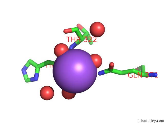
Mono view
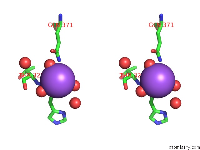
Stereo pair view
Mono view
Stereo pair view
A full contact list of Sodium with other atoms in the Na binding
site number 1 of Crystal Structure of GH1 Beta-Glucosidase TD2F2 N223G Mutant within 5.0Å range:
|
Reference:
T.Matsuzawa,
T.Jo,
T.Uchiyama,
J.A.Manninen,
T.Arakawa,
K.Miyazaki,
S.Fushinobu,
K.Yaoi.
Crystal Structure and Identification of A Key Amino Acid For Glucose Tolerance, Substrate Specificity, and Transglycosylation Activity of Metagenomic Beta-Glucosidase TD2F2 Febs J. V. 283 2340 2016.
ISSN: ISSN 1742-464X
PubMed: 27092463
DOI: 10.1111/FEBS.13743
Page generated: Mon Oct 7 19:57:50 2024
ISSN: ISSN 1742-464X
PubMed: 27092463
DOI: 10.1111/FEBS.13743
Last articles
Zn in 9MJ5Zn in 9HNW
Zn in 9G0L
Zn in 9FNE
Zn in 9DZN
Zn in 9E0I
Zn in 9D32
Zn in 9DAK
Zn in 8ZXC
Zn in 8ZUF