Sodium »
PDB 5ack-5b07 »
5afb »
Sodium in PDB 5afb: Crystal Structure of the LATROPHILIN3 Lectin and Olfactomedin Domains
Protein crystallography data
The structure of Crystal Structure of the LATROPHILIN3 Lectin and Olfactomedin Domains, PDB code: 5afb
was solved by
V.A.Jackson,
D.Del Toro,
M.Carrasquero,
P.Roversi,
K.Harlos,
R.Klein,
E.Seiradake,
with X-Ray Crystallography technique. A brief refinement statistics is given in the table below:
| Resolution Low / High (Å) | 22.91 / 2.16 |
| Space group | I 2 2 2 |
| Cell size a, b, c (Å), α, β, γ (°) | 78.430, 96.650, 101.640, 90.00, 90.00, 90.00 |
| R / Rfree (%) | 20.44 / 24.52 |
Other elements in 5afb:
The structure of Crystal Structure of the LATROPHILIN3 Lectin and Olfactomedin Domains also contains other interesting chemical elements:
| Calcium | (Ca) | 1 atom |
Sodium Binding Sites:
The binding sites of Sodium atom in the Crystal Structure of the LATROPHILIN3 Lectin and Olfactomedin Domains
(pdb code 5afb). This binding sites where shown within
5.0 Angstroms radius around Sodium atom.
In total only one binding site of Sodium was determined in the Crystal Structure of the LATROPHILIN3 Lectin and Olfactomedin Domains, PDB code: 5afb:
In total only one binding site of Sodium was determined in the Crystal Structure of the LATROPHILIN3 Lectin and Olfactomedin Domains, PDB code: 5afb:
Sodium binding site 1 out of 1 in 5afb
Go back to
Sodium binding site 1 out
of 1 in the Crystal Structure of the LATROPHILIN3 Lectin and Olfactomedin Domains
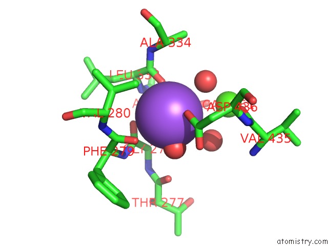
Mono view
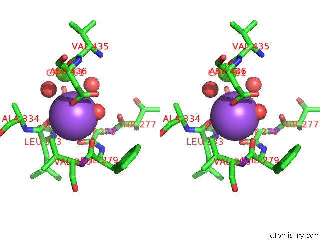
Stereo pair view
Mono view
Stereo pair view
A full contact list of Sodium with other atoms in the Na binding
site number 1 of Crystal Structure of the LATROPHILIN3 Lectin and Olfactomedin Domains within 5.0Å range:
|
Reference:
V.A.Jackson,
D.Del Toro,
M.Carrasquero,
P.Roversi,
K.Harlos,
R.Klein,
E.Seiradake.
Structural Basis of Latrophilin-Flrt Interaction. Structure 2015.
ISSN: ESSN 1878-4186
PubMed: 25728924
DOI: 10.1016/J.STR.2015.01.013
Page generated: Mon Oct 7 19:53:12 2024
ISSN: ESSN 1878-4186
PubMed: 25728924
DOI: 10.1016/J.STR.2015.01.013
Last articles
Zn in 9MJ5Zn in 9HNW
Zn in 9G0L
Zn in 9FNE
Zn in 9DZN
Zn in 9E0I
Zn in 9D32
Zn in 9DAK
Zn in 8ZXC
Zn in 8ZUF