Sodium »
PDB 4zm0-5ac7 »
4zmh »
Sodium in PDB 4zmh: Crystal Structure of A Five-Domain GH115 Alpha-Glucuronidase From the Marine Bacterium Saccharophagus Degradans 2-40T
Protein crystallography data
The structure of Crystal Structure of A Five-Domain GH115 Alpha-Glucuronidase From the Marine Bacterium Saccharophagus Degradans 2-40T, PDB code: 4zmh
was solved by
B.Nocek,
H.Cui,
W.Wang,
A.Savchenko,
with X-Ray Crystallography technique. A brief refinement statistics is given in the table below:
| Resolution Low / High (Å) | 38.15 / 1.93 |
| Space group | P 21 21 21 |
| Cell size a, b, c (Å), α, β, γ (°) | 116.442, 124.294, 180.027, 90.00, 90.00, 90.00 |
| R / Rfree (%) | 13.8 / 17 |
Sodium Binding Sites:
The binding sites of Sodium atom in the Crystal Structure of A Five-Domain GH115 Alpha-Glucuronidase From the Marine Bacterium Saccharophagus Degradans 2-40T
(pdb code 4zmh). This binding sites where shown within
5.0 Angstroms radius around Sodium atom.
In total 2 binding sites of Sodium where determined in the Crystal Structure of A Five-Domain GH115 Alpha-Glucuronidase From the Marine Bacterium Saccharophagus Degradans 2-40T, PDB code: 4zmh:
Jump to Sodium binding site number: 1; 2;
In total 2 binding sites of Sodium where determined in the Crystal Structure of A Five-Domain GH115 Alpha-Glucuronidase From the Marine Bacterium Saccharophagus Degradans 2-40T, PDB code: 4zmh:
Jump to Sodium binding site number: 1; 2;
Sodium binding site 1 out of 2 in 4zmh
Go back to
Sodium binding site 1 out
of 2 in the Crystal Structure of A Five-Domain GH115 Alpha-Glucuronidase From the Marine Bacterium Saccharophagus Degradans 2-40T
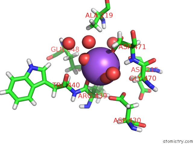
Mono view
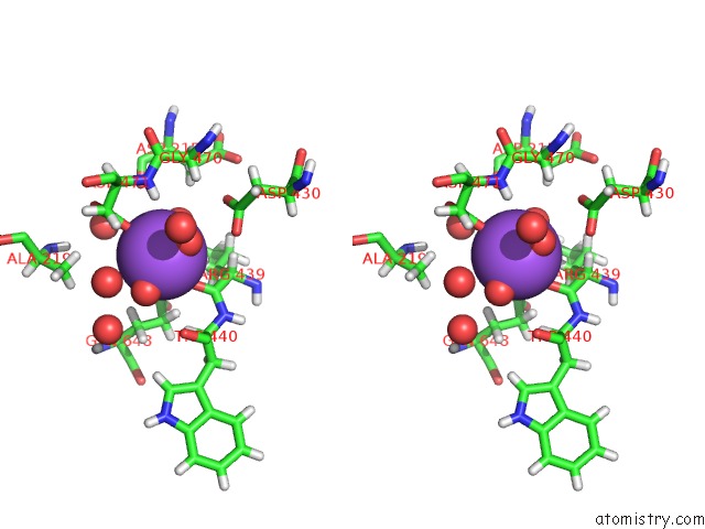
Stereo pair view
Mono view
Stereo pair view
A full contact list of Sodium with other atoms in the Na binding
site number 1 of Crystal Structure of A Five-Domain GH115 Alpha-Glucuronidase From the Marine Bacterium Saccharophagus Degradans 2-40T within 5.0Å range:
|
Sodium binding site 2 out of 2 in 4zmh
Go back to
Sodium binding site 2 out
of 2 in the Crystal Structure of A Five-Domain GH115 Alpha-Glucuronidase From the Marine Bacterium Saccharophagus Degradans 2-40T
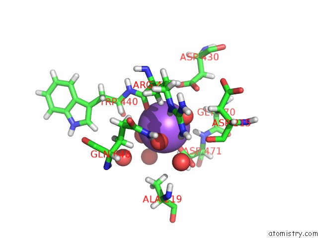
Mono view
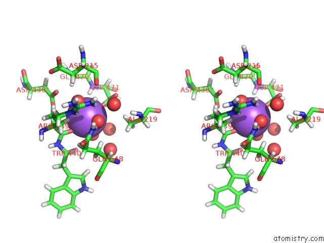
Stereo pair view
Mono view
Stereo pair view
A full contact list of Sodium with other atoms in the Na binding
site number 2 of Crystal Structure of A Five-Domain GH115 Alpha-Glucuronidase From the Marine Bacterium Saccharophagus Degradans 2-40T within 5.0Å range:
|
Reference:
W.Wang,
R.Yan,
B.P.Nocek,
T.V.Vuong,
R.Di Leo,
X.Xu,
H.Cui,
P.Gatenholm,
G.Toriz,
M.Tenkanen,
A.Savchenko,
E.R.Master.
Biochemical and Structural Characterization of A Five-Domain GH115 Alpha-Glucuronidase From the Marine Bacterium Saccharophagus Degradans 2-40T. J.Biol.Chem. V. 291 14120 2016.
ISSN: ESSN 1083-351X
PubMed: 27129264
DOI: 10.1074/JBC.M115.702944
Page generated: Mon Oct 7 19:45:07 2024
ISSN: ESSN 1083-351X
PubMed: 27129264
DOI: 10.1074/JBC.M115.702944
Last articles
Zn in 9J0NZn in 9J0O
Zn in 9J0P
Zn in 9FJX
Zn in 9EKB
Zn in 9C0F
Zn in 9CAH
Zn in 9CH0
Zn in 9CH3
Zn in 9CH1