Sodium »
PDB 4z50-4zls »
4zbk »
Sodium in PDB 4zbk: Crystal Structure of Human GGT1 in Complex with Ggstop Inhibitor
Enzymatic activity of Crystal Structure of Human GGT1 in Complex with Ggstop Inhibitor
All present enzymatic activity of Crystal Structure of Human GGT1 in Complex with Ggstop Inhibitor:
2.3.2.2; 3.4.19.13; 3.4.19.14;
2.3.2.2; 3.4.19.13; 3.4.19.14;
Protein crystallography data
The structure of Crystal Structure of Human GGT1 in Complex with Ggstop Inhibitor, PDB code: 4zbk
was solved by
S.Terzyan,
M.Hanigan,
with X-Ray Crystallography technique. A brief refinement statistics is given in the table below:
| Resolution Low / High (Å) | 47.13 / 2.18 |
| Space group | C 2 2 21 |
| Cell size a, b, c (Å), α, β, γ (°) | 105.702, 123.551, 104.189, 90.00, 90.00, 90.00 |
| R / Rfree (%) | 15.9 / 21.8 |
Other elements in 4zbk:
The structure of Crystal Structure of Human GGT1 in Complex with Ggstop Inhibitor also contains other interesting chemical elements:
| Chlorine | (Cl) | 2 atoms |
Sodium Binding Sites:
The binding sites of Sodium atom in the Crystal Structure of Human GGT1 in Complex with Ggstop Inhibitor
(pdb code 4zbk). This binding sites where shown within
5.0 Angstroms radius around Sodium atom.
In total only one binding site of Sodium was determined in the Crystal Structure of Human GGT1 in Complex with Ggstop Inhibitor, PDB code: 4zbk:
In total only one binding site of Sodium was determined in the Crystal Structure of Human GGT1 in Complex with Ggstop Inhibitor, PDB code: 4zbk:
Sodium binding site 1 out of 1 in 4zbk
Go back to
Sodium binding site 1 out
of 1 in the Crystal Structure of Human GGT1 in Complex with Ggstop Inhibitor
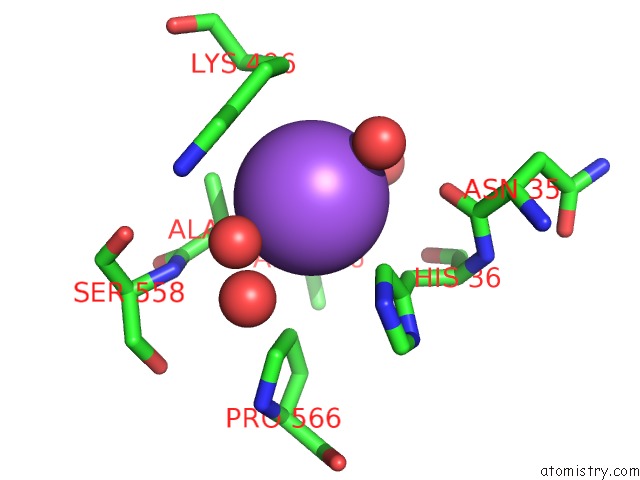
Mono view
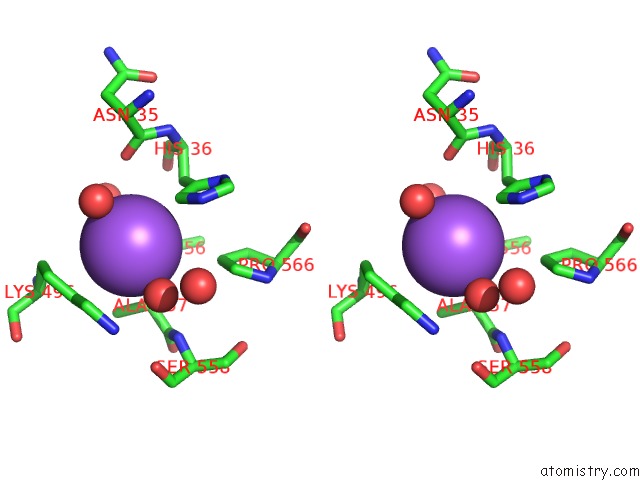
Stereo pair view
Mono view
Stereo pair view
A full contact list of Sodium with other atoms in the Na binding
site number 1 of Crystal Structure of Human GGT1 in Complex with Ggstop Inhibitor within 5.0Å range:
|
Reference:
S.S.Terzyan,
A.W.Burgett,
A.Heroux,
C.A.Smith,
B.H.Mooers,
M.H.Hanigan.
Human Gamma-Glutamyl Transpeptidase 1: Structures of the Free Enzyme, Inhibitor-Bound Tetrahedral Transition States, and Glutamate-Bound Enzyme Reveal Novel Movement Within the Active Site During Catalysis. J.Biol.Chem. V. 290 17576 2015.
ISSN: ESSN 1083-351X
PubMed: 26013825
DOI: 10.1074/JBC.M115.659680
Page generated: Mon Oct 7 19:39:56 2024
ISSN: ESSN 1083-351X
PubMed: 26013825
DOI: 10.1074/JBC.M115.659680
Last articles
F in 7QMYF in 7QMX
F in 7QMW
F in 7QMZ
F in 7QMU
F in 7QMV
F in 7QMT
F in 7QMS
F in 7QMQ
F in 7QMR