Sodium in PDB 4xmz: Crystal Structure of MET260ALA Mutant of E. Coli Aminopeptidase N in Complex with 2,4-Diaminobutyric Acid
Enzymatic activity of Crystal Structure of MET260ALA Mutant of E. Coli Aminopeptidase N in Complex with 2,4-Diaminobutyric Acid
All present enzymatic activity of Crystal Structure of MET260ALA Mutant of E. Coli Aminopeptidase N in Complex with 2,4-Diaminobutyric Acid:
3.4.11.2;
Protein crystallography data
The structure of Crystal Structure of MET260ALA Mutant of E. Coli Aminopeptidase N in Complex with 2,4-Diaminobutyric Acid, PDB code: 4xmz
was solved by
A.Addlagatta,
R.Gumpena,
with X-Ray Crystallography technique. A brief refinement statistics is given in the table below:
Resolution Low / High (Å)
|
19.81 /
2.15
|
Space group
|
P 31 2 1
|
Cell size a, b, c (Å), α, β, γ (°)
|
120.638,
120.638,
171.304,
90.00,
90.00,
120.00
|
R / Rfree (%)
|
14.3 /
18.1
|
Other elements in 4xmz:
The structure of Crystal Structure of MET260ALA Mutant of E. Coli Aminopeptidase N in Complex with 2,4-Diaminobutyric Acid also contains other interesting chemical elements:
Sodium Binding Sites:
The binding sites of Sodium atom in the Crystal Structure of MET260ALA Mutant of E. Coli Aminopeptidase N in Complex with 2,4-Diaminobutyric Acid
(pdb code 4xmz). This binding sites where shown within
5.0 Angstroms radius around Sodium atom.
In total 8 binding sites of Sodium where determined in the
Crystal Structure of MET260ALA Mutant of E. Coli Aminopeptidase N in Complex with 2,4-Diaminobutyric Acid, PDB code: 4xmz:
Jump to Sodium binding site number:
1;
2;
3;
4;
5;
6;
7;
8;
Sodium binding site 1 out
of 8 in 4xmz
Go back to
Sodium Binding Sites List in 4xmz
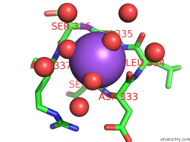
Sodium binding site 1 out
of 8 in the Crystal Structure of MET260ALA Mutant of E. Coli Aminopeptidase N in Complex with 2,4-Diaminobutyric Acid
Mono view
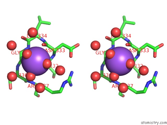
Stereo pair view
|
A full contact list of Sodium with other atoms in the Na binding
site number 1 of Crystal Structure of MET260ALA Mutant of E. Coli Aminopeptidase N in Complex with 2,4-Diaminobutyric Acid within 5.0Å range:
probe
|
atom
|
residue
|
distance (Å)
|
B
|
Occ
|
A:Na902
b:25.2
occ:1.00
|
O
|
A:HOH1878
|
2.3
|
30.8
|
1.0
|
O
|
A:GLY335
|
2.3
|
16.4
|
1.0
|
O
|
A:HOH1767
|
2.3
|
26.1
|
1.0
|
O
|
A:SER332
|
2.5
|
15.1
|
1.0
|
O
|
A:HOH1602
|
2.6
|
21.1
|
1.0
|
O
|
A:ASP333
|
2.9
|
16.4
|
1.0
|
C
|
A:GLY335
|
3.4
|
15.0
|
1.0
|
C
|
A:ASP333
|
3.4
|
16.4
|
1.0
|
C
|
A:SER332
|
3.6
|
14.5
|
1.0
|
CA
|
A:ASP333
|
3.8
|
16.0
|
1.0
|
N
|
A:GLY335
|
3.9
|
14.6
|
1.0
|
N
|
A:ARG337
|
4.0
|
18.1
|
1.0
|
O
|
A:HOH1548
|
4.1
|
28.9
|
1.0
|
N
|
A:ASP333
|
4.1
|
15.5
|
1.0
|
C
|
A:LEU334
|
4.2
|
15.8
|
1.0
|
CA
|
A:GLY335
|
4.2
|
14.6
|
1.0
|
N
|
A:LEU334
|
4.3
|
15.0
|
1.0
|
N
|
A:SER336
|
4.4
|
14.7
|
1.0
|
O
|
A:HOH1962
|
4.5
|
24.6
|
1.0
|
O
|
A:HOH1376
|
4.5
|
20.7
|
1.0
|
CA
|
A:SER336
|
4.5
|
16.7
|
1.0
|
O
|
A:HOH2075
|
4.6
|
41.0
|
1.0
|
C
|
A:SER336
|
4.6
|
17.9
|
1.0
|
CB
|
A:ARG337
|
4.6
|
18.5
|
1.0
|
O
|
A:LEU334
|
4.7
|
15.4
|
1.0
|
O
|
A:HOH1790
|
4.7
|
41.9
|
1.0
|
CA
|
A:ARG337
|
4.7
|
18.8
|
1.0
|
CA
|
A:LEU334
|
4.8
|
15.9
|
1.0
|
CA
|
A:SER332
|
4.8
|
13.0
|
1.0
|
|
Sodium binding site 2 out
of 8 in 4xmz
Go back to
Sodium Binding Sites List in 4xmz
Sodium binding site 2 out
of 8 in the Crystal Structure of MET260ALA Mutant of E. Coli Aminopeptidase N in Complex with 2,4-Diaminobutyric Acid
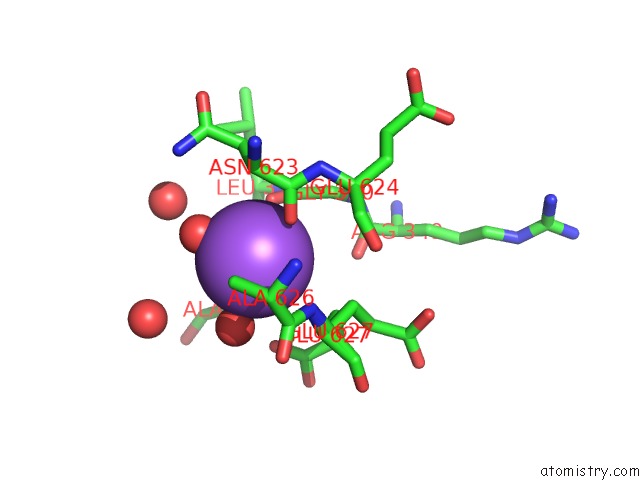
Mono view
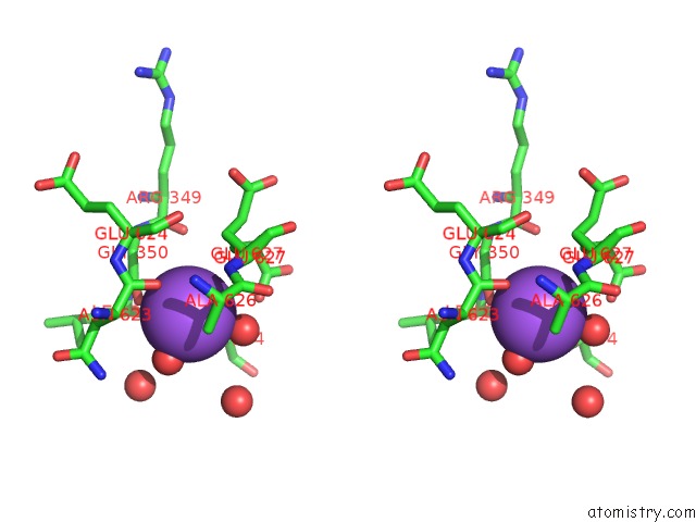
Stereo pair view
|
A full contact list of Sodium with other atoms in the Na binding
site number 2 of Crystal Structure of MET260ALA Mutant of E. Coli Aminopeptidase N in Complex with 2,4-Diaminobutyric Acid within 5.0Å range:
probe
|
atom
|
residue
|
distance (Å)
|
B
|
Occ
|
A:Na903
b:35.7
occ:1.00
|
O
|
A:HOH1866
|
2.3
|
32.3
|
1.0
|
O
|
A:ASN623
|
2.7
|
21.0
|
1.0
|
O
|
A:HOH1322
|
2.7
|
23.6
|
1.0
|
O
|
A:HOH1009
|
2.7
|
33.3
|
1.0
|
O
|
A:GLY350
|
2.8
|
26.1
|
1.0
|
N
|
A:GLU627
|
3.6
|
17.9
|
1.0
|
C
|
A:ASN623
|
3.7
|
21.2
|
1.0
|
CB
|
A:ALA354
|
3.8
|
16.0
|
1.0
|
C
|
A:GLY350
|
3.8
|
21.3
|
1.0
|
CB
|
A:GLU627
|
3.8
|
20.3
|
0.5
|
CB
|
A:ALA626
|
3.8
|
18.5
|
1.0
|
OE2
|
A:GLU627
|
3.9
|
18.2
|
0.5
|
CA
|
A:GLU627
|
4.1
|
19.2
|
0.5
|
CA
|
A:GLU627
|
4.1
|
18.4
|
0.5
|
CA
|
A:ASN623
|
4.1
|
23.9
|
1.0
|
CB
|
A:GLU627
|
4.1
|
18.0
|
0.5
|
C
|
A:ALA626
|
4.2
|
18.0
|
1.0
|
CA
|
A:GLY350
|
4.3
|
19.6
|
1.0
|
O
|
A:HOH1750
|
4.4
|
30.6
|
1.0
|
CB
|
A:ASN623
|
4.5
|
26.1
|
1.0
|
CA
|
A:ALA626
|
4.5
|
17.4
|
1.0
|
CA
|
A:ALA354
|
4.7
|
16.6
|
1.0
|
CG
|
A:GLU627
|
4.7
|
21.3
|
0.5
|
O
|
A:ARG349
|
4.7
|
23.0
|
1.0
|
CD
|
A:GLU627
|
4.7
|
21.9
|
0.5
|
N
|
A:GLU624
|
4.7
|
19.4
|
1.0
|
O
|
A:HOH1834
|
4.7
|
18.5
|
1.0
|
N
|
A:LEU351
|
4.8
|
18.6
|
1.0
|
N
|
A:ALA354
|
4.8
|
15.4
|
1.0
|
N
|
A:ALA626
|
4.9
|
16.9
|
1.0
|
O
|
A:ALA626
|
5.0
|
19.7
|
1.0
|
|
Sodium binding site 3 out
of 8 in 4xmz
Go back to
Sodium Binding Sites List in 4xmz
Sodium binding site 3 out
of 8 in the Crystal Structure of MET260ALA Mutant of E. Coli Aminopeptidase N in Complex with 2,4-Diaminobutyric Acid
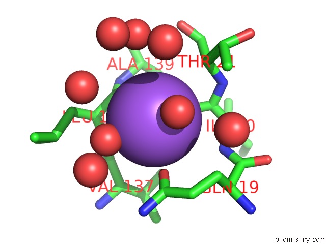
Mono view
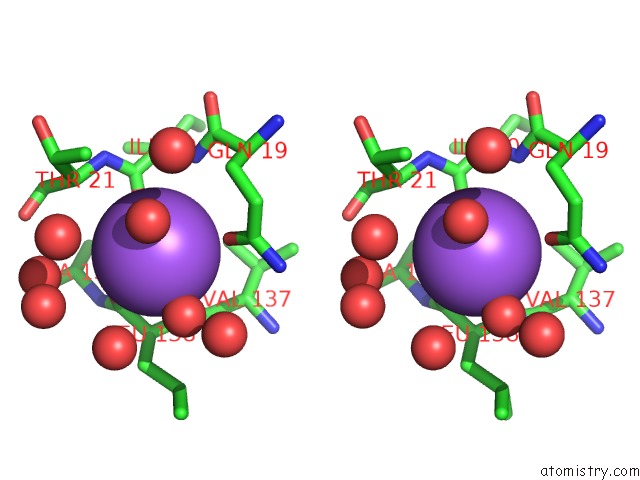
Stereo pair view
|
A full contact list of Sodium with other atoms in the Na binding
site number 3 of Crystal Structure of MET260ALA Mutant of E. Coli Aminopeptidase N in Complex with 2,4-Diaminobutyric Acid within 5.0Å range:
probe
|
atom
|
residue
|
distance (Å)
|
B
|
Occ
|
A:Na904
b:34.6
occ:1.00
|
O
|
A:HOH1883
|
2.6
|
33.2
|
1.0
|
O
|
A:LEU138
|
2.6
|
15.9
|
1.0
|
OE1
|
A:GLN19
|
2.7
|
21.0
|
1.0
|
O
|
A:HOH2044
|
2.8
|
29.9
|
1.0
|
O
|
A:ILE20
|
3.0
|
19.3
|
1.0
|
CD
|
A:GLN19
|
3.5
|
18.1
|
1.0
|
C
|
A:LEU138
|
3.6
|
15.5
|
1.0
|
N
|
A:LEU138
|
3.7
|
14.6
|
1.0
|
C
|
A:ILE20
|
3.8
|
18.0
|
1.0
|
O
|
A:HOH1979
|
4.0
|
30.2
|
1.0
|
CB
|
A:GLN19
|
4.0
|
17.2
|
1.0
|
CG
|
A:GLN19
|
4.0
|
17.2
|
1.0
|
CA
|
A:THR21
|
4.1
|
17.7
|
1.0
|
CA
|
A:LEU138
|
4.1
|
16.2
|
1.0
|
O
|
A:HOH1319
|
4.2
|
21.7
|
1.0
|
N
|
A:THR21
|
4.3
|
16.7
|
1.0
|
CG2
|
A:THR21
|
4.3
|
17.6
|
1.0
|
N
|
A:ILE20
|
4.3
|
17.4
|
1.0
|
NE2
|
A:GLN19
|
4.3
|
18.1
|
1.0
|
CB
|
A:LEU138
|
4.5
|
17.8
|
1.0
|
O
|
A:HOH1935
|
4.5
|
12.2
|
0.5
|
O
|
A:HOH1951
|
4.6
|
31.4
|
1.0
|
C
|
A:VAL137
|
4.6
|
14.9
|
1.0
|
N
|
A:ALA139
|
4.7
|
15.2
|
1.0
|
CA
|
A:ILE20
|
4.8
|
17.3
|
1.0
|
CB
|
A:THR21
|
4.8
|
17.9
|
1.0
|
CA
|
A:VAL137
|
4.8
|
14.4
|
1.0
|
O
|
A:HOH2102
|
4.9
|
35.0
|
1.0
|
CA
|
A:GLN19
|
4.9
|
17.3
|
1.0
|
O
|
A:HOH1333
|
4.9
|
15.2
|
1.0
|
C
|
A:GLN19
|
5.0
|
17.4
|
1.0
|
|
Sodium binding site 4 out
of 8 in 4xmz
Go back to
Sodium Binding Sites List in 4xmz
Sodium binding site 4 out
of 8 in the Crystal Structure of MET260ALA Mutant of E. Coli Aminopeptidase N in Complex with 2,4-Diaminobutyric Acid
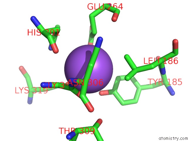
Mono view
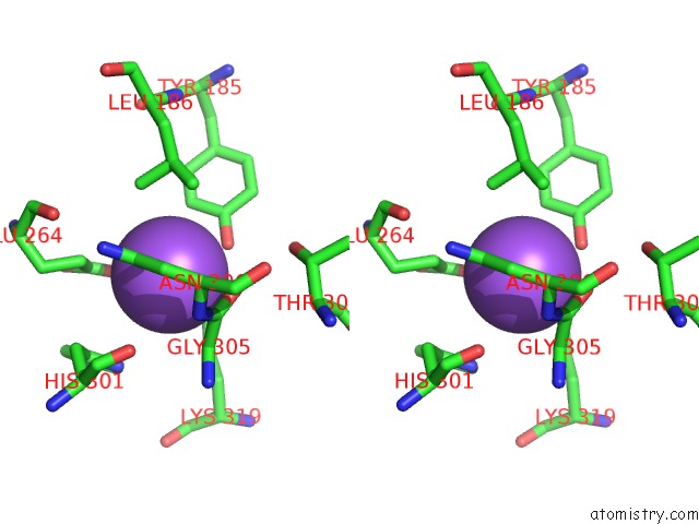
Stereo pair view
|
A full contact list of Sodium with other atoms in the Na binding
site number 4 of Crystal Structure of MET260ALA Mutant of E. Coli Aminopeptidase N in Complex with 2,4-Diaminobutyric Acid within 5.0Å range:
probe
|
atom
|
residue
|
distance (Å)
|
B
|
Occ
|
A:Na905
b:23.8
occ:1.00
|
OH
|
A:TYR185
|
2.7
|
10.7
|
1.0
|
OE2
|
A:GLU264
|
2.8
|
17.3
|
1.0
|
OD1
|
A:ASN306
|
2.8
|
15.9
|
1.0
|
O
|
A:GLY305
|
3.1
|
14.0
|
1.0
|
C
|
A:GLY305
|
3.2
|
14.2
|
1.0
|
CA
|
A:GLY305
|
3.4
|
13.4
|
1.0
|
CD
|
A:LYS319
|
3.4
|
12.8
|
1.0
|
CG
|
A:GLU264
|
3.5
|
14.6
|
1.0
|
CE1
|
A:TYR185
|
3.5
|
12.3
|
1.0
|
CZ
|
A:TYR185
|
3.5
|
12.3
|
1.0
|
CD
|
A:GLU264
|
3.6
|
15.9
|
1.0
|
CE
|
A:LYS319
|
3.7
|
12.9
|
1.0
|
CD2
|
A:LEU186
|
3.8
|
13.7
|
1.0
|
N
|
A:ASN306
|
3.9
|
12.8
|
1.0
|
CG
|
A:ASN306
|
4.0
|
13.7
|
1.0
|
NZ
|
A:LYS319
|
4.0
|
12.4
|
1.0
|
ND1
|
A:HIS301
|
4.1
|
11.9
|
1.0
|
CD1
|
A:LEU186
|
4.3
|
14.6
|
1.0
|
CA
|
A:ASN306
|
4.4
|
12.9
|
1.0
|
CG
|
A:LEU186
|
4.5
|
13.8
|
1.0
|
CG
|
A:HIS301
|
4.5
|
12.8
|
1.0
|
CB
|
A:HIS301
|
4.7
|
12.2
|
1.0
|
O
|
A:HIS301
|
4.7
|
12.2
|
1.0
|
CE1
|
A:HIS301
|
4.7
|
12.9
|
1.0
|
N
|
A:GLY305
|
4.7
|
13.5
|
1.0
|
CG
|
A:LYS319
|
4.7
|
13.2
|
1.0
|
OE1
|
A:GLU264
|
4.8
|
15.2
|
1.0
|
CB
|
A:ASN306
|
4.8
|
13.9
|
1.0
|
CD1
|
A:TYR185
|
4.9
|
12.8
|
1.0
|
CE2
|
A:TYR185
|
4.9
|
12.2
|
1.0
|
ND2
|
A:ASN306
|
4.9
|
14.1
|
1.0
|
CB
|
A:GLU264
|
4.9
|
13.8
|
1.0
|
OG1
|
A:THR309
|
4.9
|
15.1
|
1.0
|
|
Sodium binding site 5 out
of 8 in 4xmz
Go back to
Sodium Binding Sites List in 4xmz
Sodium binding site 5 out
of 8 in the Crystal Structure of MET260ALA Mutant of E. Coli Aminopeptidase N in Complex with 2,4-Diaminobutyric Acid
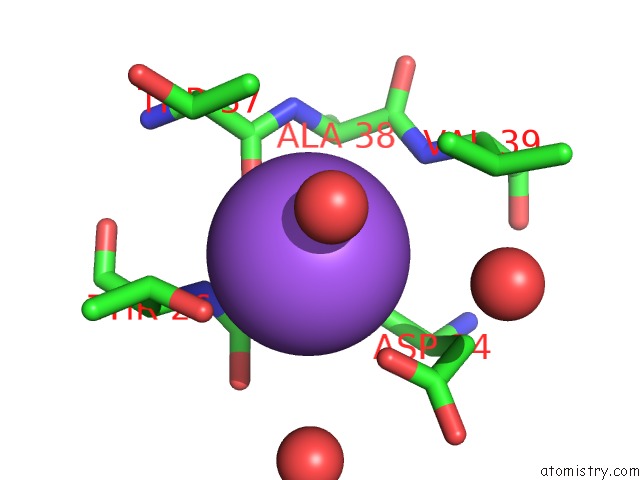
Mono view
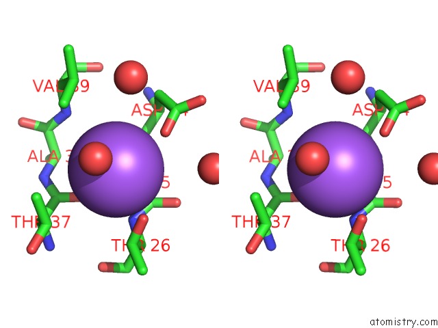
Stereo pair view
|
A full contact list of Sodium with other atoms in the Na binding
site number 5 of Crystal Structure of MET260ALA Mutant of E. Coli Aminopeptidase N in Complex with 2,4-Diaminobutyric Acid within 5.0Å range:
probe
|
atom
|
residue
|
distance (Å)
|
B
|
Occ
|
A:Na906
b:33.6
occ:1.00
|
OG1
|
A:THR26
|
2.7
|
16.0
|
1.0
|
O
|
A:HOH1982
|
2.8
|
38.2
|
1.0
|
OD1
|
A:ASP24
|
2.9
|
40.7
|
1.0
|
O
|
A:ASP24
|
3.0
|
17.4
|
1.0
|
O
|
A:THR37
|
3.1
|
19.7
|
1.0
|
N
|
A:THR26
|
3.3
|
15.2
|
1.0
|
C
|
A:LEU25
|
3.4
|
16.7
|
1.0
|
C
|
A:ASP24
|
3.5
|
17.4
|
1.0
|
CB
|
A:THR26
|
3.6
|
17.8
|
1.0
|
CA
|
A:LEU25
|
3.7
|
15.7
|
1.0
|
N
|
A:LEU25
|
3.8
|
14.2
|
1.0
|
C
|
A:THR37
|
3.9
|
18.5
|
1.0
|
O
|
A:LEU25
|
3.9
|
16.6
|
1.0
|
CG2
|
A:VAL39
|
4.0
|
17.4
|
1.0
|
CG2
|
A:THR37
|
4.0
|
20.8
|
1.0
|
CG
|
A:ASP24
|
4.0
|
32.5
|
1.0
|
CA
|
A:THR26
|
4.0
|
16.2
|
1.0
|
CB
|
A:THR37
|
4.2
|
18.3
|
1.0
|
CB
|
A:ASP24
|
4.4
|
24.9
|
1.0
|
N
|
A:VAL39
|
4.4
|
13.7
|
1.0
|
N
|
A:ALA38
|
4.5
|
15.7
|
1.0
|
CA
|
A:ASP24
|
4.5
|
17.9
|
1.0
|
CA
|
A:ALA38
|
4.6
|
16.3
|
1.0
|
O
|
A:HOH1554
|
4.6
|
21.2
|
1.0
|
CA
|
A:THR37
|
4.7
|
16.8
|
1.0
|
CB
|
A:VAL39
|
4.7
|
17.9
|
1.0
|
O
|
A:HOH1895
|
4.8
|
39.3
|
1.0
|
CG2
|
A:THR26
|
4.9
|
19.4
|
1.0
|
C
|
A:ALA38
|
4.9
|
15.5
|
1.0
|
|
Sodium binding site 6 out
of 8 in 4xmz
Go back to
Sodium Binding Sites List in 4xmz
Sodium binding site 6 out
of 8 in the Crystal Structure of MET260ALA Mutant of E. Coli Aminopeptidase N in Complex with 2,4-Diaminobutyric Acid
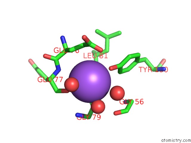
Mono view
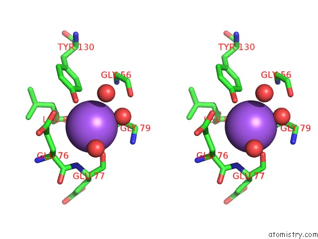
Stereo pair view
|
A full contact list of Sodium with other atoms in the Na binding
site number 6 of Crystal Structure of MET260ALA Mutant of E. Coli Aminopeptidase N in Complex with 2,4-Diaminobutyric Acid within 5.0Å range:
probe
|
atom
|
residue
|
distance (Å)
|
B
|
Occ
|
A:Na907
b:35.8
occ:1.00
|
O
|
A:HOH1837
|
2.6
|
34.8
|
1.0
|
OH
|
A:TYR130
|
2.7
|
26.9
|
1.0
|
O
|
A:HOH1411
|
2.8
|
28.5
|
1.0
|
O
|
A:ALA80
|
3.0
|
16.4
|
1.0
|
O
|
A:GLU77
|
3.0
|
23.6
|
1.0
|
N
|
A:ALA80
|
3.6
|
19.3
|
1.0
|
CZ
|
A:TYR130
|
3.7
|
22.0
|
1.0
|
O
|
A:HOH1545
|
3.7
|
28.0
|
1.0
|
C
|
A:ALA80
|
3.8
|
18.5
|
1.0
|
CE1
|
A:TYR130
|
3.8
|
19.8
|
1.0
|
CB
|
A:GLU76
|
3.9
|
25.8
|
1.0
|
OE2
|
A:GLU76
|
3.9
|
27.3
|
1.0
|
CG
|
A:GLU76
|
4.0
|
24.7
|
1.0
|
C
|
A:GLU77
|
4.1
|
26.5
|
1.0
|
N
|
A:GLU77
|
4.1
|
26.7
|
1.0
|
CD
|
A:GLU76
|
4.2
|
25.1
|
1.0
|
CA
|
A:ALA80
|
4.2
|
18.3
|
1.0
|
N
|
A:GLY79
|
4.3
|
23.4
|
1.0
|
C
|
A:GLY79
|
4.4
|
21.1
|
1.0
|
CA
|
A:GLU76
|
4.4
|
25.5
|
1.0
|
CA
|
A:GLY79
|
4.6
|
24.2
|
1.0
|
C
|
A:GLU76
|
4.6
|
27.1
|
1.0
|
O
|
A:GLY56
|
4.7
|
17.7
|
1.0
|
N
|
A:LEU81
|
4.8
|
16.8
|
1.0
|
CA
|
A:GLU77
|
4.8
|
27.0
|
1.0
|
|
Sodium binding site 7 out
of 8 in 4xmz
Go back to
Sodium Binding Sites List in 4xmz
Sodium binding site 7 out
of 8 in the Crystal Structure of MET260ALA Mutant of E. Coli Aminopeptidase N in Complex with 2,4-Diaminobutyric Acid
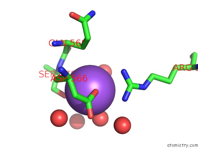
Mono view
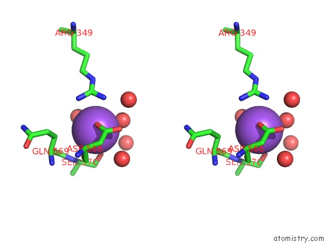
Stereo pair view
|
A full contact list of Sodium with other atoms in the Na binding
site number 7 of Crystal Structure of MET260ALA Mutant of E. Coli Aminopeptidase N in Complex with 2,4-Diaminobutyric Acid within 5.0Å range:
probe
|
atom
|
residue
|
distance (Å)
|
B
|
Occ
|
A:Na908
b:31.5
occ:1.00
|
OD1
|
A:ASP566
|
2.6
|
17.0
|
1.0
|
OG
|
A:SER570
|
2.7
|
16.0
|
1.0
|
O
|
A:HOH1955
|
2.8
|
28.5
|
1.0
|
O
|
A:HOH2005
|
3.0
|
29.3
|
1.0
|
O
|
A:ASP566
|
3.2
|
16.0
|
1.0
|
CG
|
A:ASP566
|
3.4
|
18.1
|
1.0
|
NH2
|
A:ARG349
|
3.6
|
26.4
|
1.0
|
NH1
|
A:ARG349
|
3.7
|
27.9
|
1.0
|
N
|
A:SER570
|
3.7
|
14.5
|
1.0
|
O
|
A:HOH1581
|
3.8
|
31.2
|
1.0
|
CZ
|
A:ARG349
|
3.9
|
25.6
|
1.0
|
CB
|
A:SER570
|
3.9
|
15.0
|
1.0
|
CB
|
A:GLN569
|
4.0
|
16.9
|
1.0
|
OD2
|
A:ASP566
|
4.0
|
22.2
|
1.0
|
CA
|
A:SER570
|
4.1
|
15.2
|
1.0
|
C
|
A:ASP566
|
4.1
|
16.9
|
1.0
|
C
|
A:GLN569
|
4.3
|
16.6
|
1.0
|
O
|
A:HOH1031
|
4.3
|
32.5
|
1.0
|
O
|
A:HOH1564
|
4.4
|
39.2
|
1.0
|
CA
|
A:ASP566
|
4.4
|
15.8
|
1.0
|
O
|
A:HOH2061
|
4.5
|
30.4
|
1.0
|
CB
|
A:ASP566
|
4.5
|
16.1
|
1.0
|
CA
|
A:GLN569
|
4.7
|
16.3
|
1.0
|
NE
|
A:ARG349
|
4.8
|
24.8
|
1.0
|
CG
|
A:GLN569
|
4.9
|
17.0
|
1.0
|
O
|
A:GLN569
|
5.0
|
15.2
|
1.0
|
|
Sodium binding site 8 out
of 8 in 4xmz
Go back to
Sodium Binding Sites List in 4xmz
Sodium binding site 8 out
of 8 in the Crystal Structure of MET260ALA Mutant of E. Coli Aminopeptidase N in Complex with 2,4-Diaminobutyric Acid
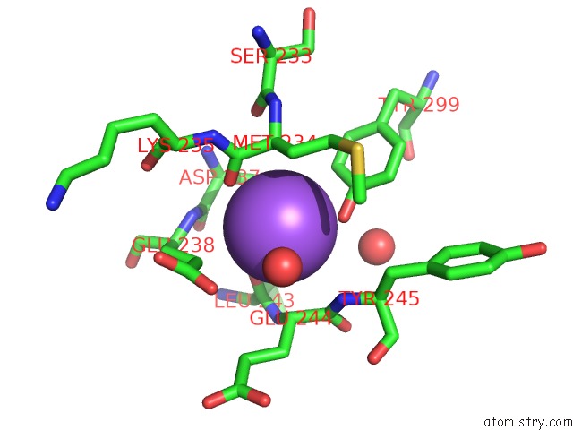
Mono view
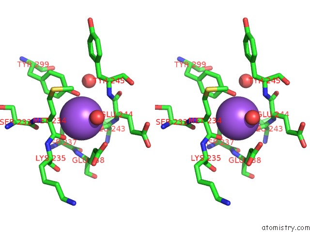
Stereo pair view
|
A full contact list of Sodium with other atoms in the Na binding
site number 8 of Crystal Structure of MET260ALA Mutant of E. Coli Aminopeptidase N in Complex with 2,4-Diaminobutyric Acid within 5.0Å range:
probe
|
atom
|
residue
|
distance (Å)
|
B
|
Occ
|
A:Na909
b:19.8
occ:1.00
|
OH
|
A:TYR299
|
2.6
|
14.3
|
1.0
|
O
|
A:HOH1479
|
2.7
|
19.1
|
1.0
|
O
|
A:LEU243
|
2.7
|
15.2
|
1.0
|
O
|
A:MET234
|
2.8
|
14.4
|
1.0
|
CZ
|
A:TYR299
|
3.4
|
15.0
|
1.0
|
CE2
|
A:TYR299
|
3.5
|
14.5
|
1.0
|
C
|
A:MET234
|
3.6
|
14.1
|
1.0
|
CA
|
A:MET234
|
3.6
|
15.0
|
1.0
|
CA
|
A:GLU244
|
3.6
|
14.4
|
1.0
|
CG
|
A:GLU238
|
3.6
|
16.8
|
1.0
|
C
|
A:LEU243
|
3.7
|
15.2
|
1.0
|
CB
|
A:ASP237
|
3.8
|
13.6
|
1.0
|
N
|
A:TYR245
|
3.8
|
13.1
|
1.0
|
C
|
A:GLU244
|
4.0
|
13.4
|
1.0
|
N
|
A:GLU238
|
4.0
|
14.8
|
1.0
|
CB
|
A:MET234
|
4.0
|
16.4
|
1.0
|
N
|
A:GLU244
|
4.1
|
14.1
|
1.0
|
O
|
A:HOH1414
|
4.4
|
14.2
|
1.0
|
C
|
A:ASP237
|
4.4
|
14.5
|
1.0
|
OE2
|
A:GLU238
|
4.5
|
20.3
|
1.0
|
CD
|
A:GLU238
|
4.5
|
18.6
|
1.0
|
CA
|
A:ASP237
|
4.5
|
13.1
|
1.0
|
CB
|
A:GLU238
|
4.6
|
16.6
|
1.0
|
CA
|
A:GLU238
|
4.6
|
14.9
|
1.0
|
CE1
|
A:TYR299
|
4.7
|
13.4
|
1.0
|
CG
|
A:MET234
|
4.8
|
17.9
|
1.0
|
O
|
A:SER233
|
4.8
|
15.1
|
1.0
|
N
|
A:LYS235
|
4.8
|
14.6
|
1.0
|
CD2
|
A:TYR299
|
4.8
|
13.5
|
1.0
|
CG
|
A:ASP237
|
4.8
|
14.4
|
1.0
|
N
|
A:ASP237
|
4.8
|
12.7
|
1.0
|
CA
|
A:TYR245
|
4.9
|
13.9
|
1.0
|
OD2
|
A:ASP237
|
4.9
|
13.7
|
1.0
|
O
|
A:GLU244
|
4.9
|
12.5
|
1.0
|
CB
|
A:GLU244
|
4.9
|
15.6
|
1.0
|
N
|
A:MET234
|
5.0
|
15.1
|
1.0
|
CA
|
A:LEU243
|
5.0
|
14.7
|
1.0
|
|
Reference:
A.Addlagatta,
R.Gumpena.
Crystal Structure of MET260ALA Mutant of E. Coli Aminopeptidase N in Complex with 2,4-Diaminobutyric Acid To Be Published.
Page generated: Mon Oct 7 19:11:31 2024
|