Sodium »
PDB 4q3d-4qom »
4q7w »
Sodium in PDB 4q7w: Crystal Structure of 1-Hydroxy-6-Methylpyridine-2(1H)-Thione Bound to Human Carbonic Anhydrase II
Enzymatic activity of Crystal Structure of 1-Hydroxy-6-Methylpyridine-2(1H)-Thione Bound to Human Carbonic Anhydrase II
All present enzymatic activity of Crystal Structure of 1-Hydroxy-6-Methylpyridine-2(1H)-Thione Bound to Human Carbonic Anhydrase II:
4.2.1.1;
4.2.1.1;
Protein crystallography data
The structure of Crystal Structure of 1-Hydroxy-6-Methylpyridine-2(1H)-Thione Bound to Human Carbonic Anhydrase II, PDB code: 4q7w
was solved by
D.P.Martin,
S.M.Cohen,
with X-Ray Crystallography technique. A brief refinement statistics is given in the table below:
| Resolution Low / High (Å) | 30.60 / 1.45 |
| Space group | P 1 21 1 |
| Cell size a, b, c (Å), α, β, γ (°) | 42.189, 41.418, 72.173, 90.00, 104.50, 90.00 |
| R / Rfree (%) | 17.9 / 21.1 |
Other elements in 4q7w:
The structure of Crystal Structure of 1-Hydroxy-6-Methylpyridine-2(1H)-Thione Bound to Human Carbonic Anhydrase II also contains other interesting chemical elements:
| Mercury | (Hg) | 1 atom |
| Zinc | (Zn) | 1 atom |
Sodium Binding Sites:
The binding sites of Sodium atom in the Crystal Structure of 1-Hydroxy-6-Methylpyridine-2(1H)-Thione Bound to Human Carbonic Anhydrase II
(pdb code 4q7w). This binding sites where shown within
5.0 Angstroms radius around Sodium atom.
In total only one binding site of Sodium was determined in the Crystal Structure of 1-Hydroxy-6-Methylpyridine-2(1H)-Thione Bound to Human Carbonic Anhydrase II, PDB code: 4q7w:
In total only one binding site of Sodium was determined in the Crystal Structure of 1-Hydroxy-6-Methylpyridine-2(1H)-Thione Bound to Human Carbonic Anhydrase II, PDB code: 4q7w:
Sodium binding site 1 out of 1 in 4q7w
Go back to
Sodium binding site 1 out
of 1 in the Crystal Structure of 1-Hydroxy-6-Methylpyridine-2(1H)-Thione Bound to Human Carbonic Anhydrase II
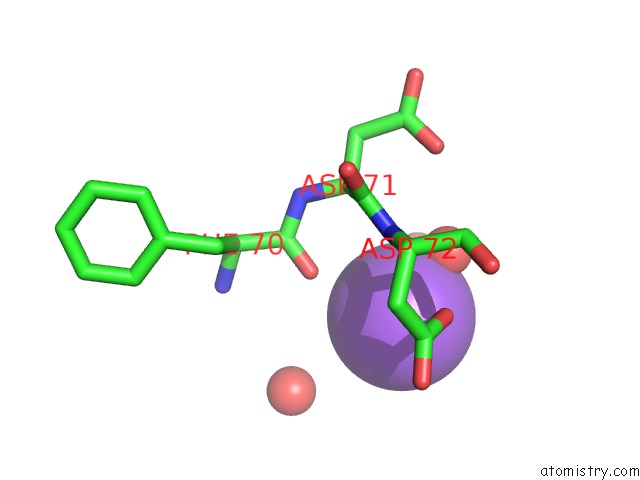
Mono view
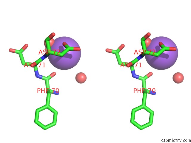
Stereo pair view
Mono view
Stereo pair view
A full contact list of Sodium with other atoms in the Na binding
site number 1 of Crystal Structure of 1-Hydroxy-6-Methylpyridine-2(1H)-Thione Bound to Human Carbonic Anhydrase II within 5.0Å range:
|
Reference:
D.P.Martin,
P.G.Blachly,
J.A.Mccammon,
S.M.Cohen.
Exploring the Influence of the Protein Environment on Metal-Binding Pharmacophores. J.Med.Chem. V. 57 7126 2014.
ISSN: ISSN 0022-2623
PubMed: 25116076
DOI: 10.1021/JM500984B
Page generated: Mon Oct 7 17:57:05 2024
ISSN: ISSN 0022-2623
PubMed: 25116076
DOI: 10.1021/JM500984B
Last articles
Zn in 9MJ5Zn in 9HNW
Zn in 9G0L
Zn in 9FNE
Zn in 9DZN
Zn in 9E0I
Zn in 9D32
Zn in 9DAK
Zn in 8ZXC
Zn in 8ZUF