Sodium »
PDB 4q3d-4qom »
4q4i »
Sodium in PDB 4q4i: Crystal Structure of E.Coli Aminopeptidase N in Complex with Amastatin
Enzymatic activity of Crystal Structure of E.Coli Aminopeptidase N in Complex with Amastatin
All present enzymatic activity of Crystal Structure of E.Coli Aminopeptidase N in Complex with Amastatin:
3.4.11.2;
3.4.11.2;
Protein crystallography data
The structure of Crystal Structure of E.Coli Aminopeptidase N in Complex with Amastatin, PDB code: 4q4i
was solved by
R.Reddi,
R.J.Ganji,
A.Addlagatta,
with X-Ray Crystallography technique. A brief refinement statistics is given in the table below:
| Resolution Low / High (Å) | 49.72 / 2.31 |
| Space group | P 31 2 1 |
| Cell size a, b, c (Å), α, β, γ (°) | 119.386, 119.386, 170.129, 90.00, 90.00, 120.00 |
| R / Rfree (%) | 14 / 17.9 |
Other elements in 4q4i:
The structure of Crystal Structure of E.Coli Aminopeptidase N in Complex with Amastatin also contains other interesting chemical elements:
| Zinc | (Zn) | 1 atom |
Sodium Binding Sites:
The binding sites of Sodium atom in the Crystal Structure of E.Coli Aminopeptidase N in Complex with Amastatin
(pdb code 4q4i). This binding sites where shown within
5.0 Angstroms radius around Sodium atom.
In total 3 binding sites of Sodium where determined in the Crystal Structure of E.Coli Aminopeptidase N in Complex with Amastatin, PDB code: 4q4i:
Jump to Sodium binding site number: 1; 2; 3;
In total 3 binding sites of Sodium where determined in the Crystal Structure of E.Coli Aminopeptidase N in Complex with Amastatin, PDB code: 4q4i:
Jump to Sodium binding site number: 1; 2; 3;
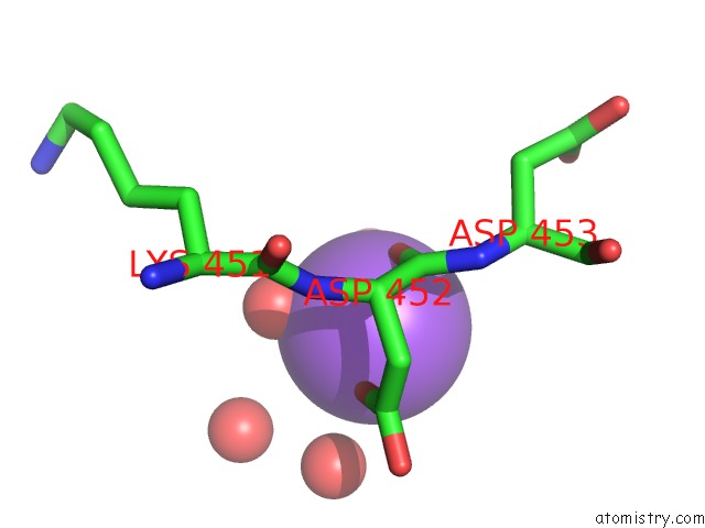
Sodium binding site 1 out of 3 in 4q4i
Go back to
Sodium binding site 1 out
of 3 in the Crystal Structure of E.Coli Aminopeptidase N in Complex with Amastatin

Mono view

Stereo pair view
Mono view
Stereo pair view
A full contact list of Sodium with other atoms in the Na binding
site number 1 of Crystal Structure of E.Coli Aminopeptidase N in Complex with Amastatin within 5.0Å range:
|
Sodium binding site 2 out of 3 in 4q4i
Go back to
Sodium binding site 2 out
of 3 in the Crystal Structure of E.Coli Aminopeptidase N in Complex with Amastatin
Mono view

Stereo pair view
Mono view
Stereo pair view
A full contact list of Sodium with other atoms in the Na binding
site number 2 of Crystal Structure of E.Coli Aminopeptidase N in Complex with Amastatin within 5.0Å range:
|
Sodium binding site 3 out of 3 in 4q4i
Go back to
Sodium binding site 3 out
of 3 in the Crystal Structure of E.Coli Aminopeptidase N in Complex with Amastatin

Mono view

Stereo pair view
Mono view
Stereo pair view
A full contact list of Sodium with other atoms in the Na binding
site number 3 of Crystal Structure of E.Coli Aminopeptidase N in Complex with Amastatin within 5.0Å range:
|
Reference:
R.J.Ganji,
R.Reddi,
R.Gumpena,
A.K.Marapaka,
T.Arya,
P.Sankoju,
S.Bhukya,
A.Addlagatta.
Structural Basis For the Inhibition of M1 Family Aminopeptidases By the Natural Product Actinonin: Crystal Structure in Complex with E. Coli Aminopeptidase N Protein Sci. 2015.
ISSN: ESSN 1469-896X
PubMed: 25644575
DOI: 10.1002/PRO.2653
Page generated: Mon Oct 7 17:55:53 2024
ISSN: ESSN 1469-896X
PubMed: 25644575
DOI: 10.1002/PRO.2653
Last articles
Cl in 5IDYCl in 5IEQ
Cl in 5IDN
Cl in 5ICR
Cl in 5IBP
Cl in 5ICU
Cl in 5ICP
Cl in 5IA3
Cl in 5IAE
Cl in 5IBN