Sodium »
PDB 4ppf-4q3c »
4pyk »
Sodium in PDB 4pyk: Human Comt, Double Domain Swap
Enzymatic activity of Human Comt, Double Domain Swap
All present enzymatic activity of Human Comt, Double Domain Swap:
2.1.1.6;
2.1.1.6;
Protein crystallography data
The structure of Human Comt, Double Domain Swap, PDB code: 4pyk
was solved by
A.Ehler,
J.Benz,
D.Schlatter,
M.G.Rudolph,
with X-Ray Crystallography technique. A brief refinement statistics is given in the table below:
| Resolution Low / High (Å) | 40.22 / 2.22 |
| Space group | C 2 2 21 |
| Cell size a, b, c (Å), α, β, γ (°) | 54.786, 66.825, 128.032, 90.00, 90.00, 90.00 |
| R / Rfree (%) | 17.2 / 24.3 |
Other elements in 4pyk:
The structure of Human Comt, Double Domain Swap also contains other interesting chemical elements:
| Magnesium | (Mg) | 1 atom |
| Chlorine | (Cl) | 1 atom |
Sodium Binding Sites:
The binding sites of Sodium atom in the Human Comt, Double Domain Swap
(pdb code 4pyk). This binding sites where shown within
5.0 Angstroms radius around Sodium atom.
In total only one binding site of Sodium was determined in the Human Comt, Double Domain Swap, PDB code: 4pyk:
In total only one binding site of Sodium was determined in the Human Comt, Double Domain Swap, PDB code: 4pyk:
Sodium binding site 1 out of 1 in 4pyk
Go back to
Sodium binding site 1 out
of 1 in the Human Comt, Double Domain Swap
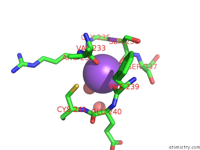
Mono view
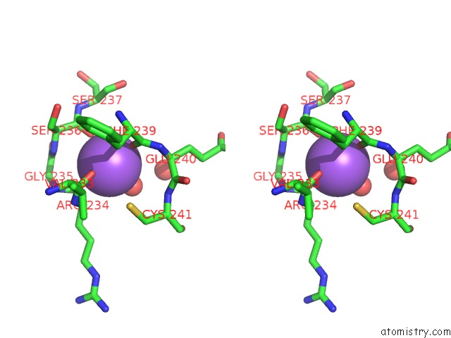
Stereo pair view
Mono view
Stereo pair view
A full contact list of Sodium with other atoms in the Na binding
site number 1 of Human Comt, Double Domain Swap within 5.0Å range:
|
Reference:
A.Ehler,
J.Benz,
D.Schlatter,
M.G.Rudolph.
Mapping the Conformational Space Accessible to Catechol-O-Methyltransferase. Acta Crystallogr.,Sect.D V. 70 2163 2014.
ISSN: ISSN 0907-4449
PubMed: 25084335
DOI: 10.1107/S1399004714012917
Page generated: Mon Oct 7 17:52:18 2024
ISSN: ISSN 0907-4449
PubMed: 25084335
DOI: 10.1107/S1399004714012917
Last articles
Zn in 9J0NZn in 9J0O
Zn in 9J0P
Zn in 9FJX
Zn in 9EKB
Zn in 9C0F
Zn in 9CAH
Zn in 9CH0
Zn in 9CH3
Zn in 9CH1