Sodium »
PDB 4pd8-4pp4 »
4pmo »
Sodium in PDB 4pmo: Crystal Structure of the Mycobacterium Tuberculosis Tat-Secreted Protein RV2525C, Monoclinic Crystal Form I
Protein crystallography data
The structure of Crystal Structure of the Mycobacterium Tuberculosis Tat-Secreted Protein RV2525C, Monoclinic Crystal Form I, PDB code: 4pmo
was solved by
M.Bellinzoni,
A.Haouz,
W.Shepard,
P.M.Alzari,
with X-Ray Crystallography technique. A brief refinement statistics is given in the table below:
| Resolution Low / High (Å) | 36.95 / 1.33 |
| Space group | C 1 2 1 |
| Cell size a, b, c (Å), α, β, γ (°) | 99.442, 73.679, 74.415, 90.00, 130.85, 90.00 |
| R / Rfree (%) | 15 / 16.5 |
Sodium Binding Sites:
The binding sites of Sodium atom in the Crystal Structure of the Mycobacterium Tuberculosis Tat-Secreted Protein RV2525C, Monoclinic Crystal Form I
(pdb code 4pmo). This binding sites where shown within
5.0 Angstroms radius around Sodium atom.
In total 6 binding sites of Sodium where determined in the Crystal Structure of the Mycobacterium Tuberculosis Tat-Secreted Protein RV2525C, Monoclinic Crystal Form I, PDB code: 4pmo:
Jump to Sodium binding site number: 1; 2; 3; 4; 5; 6;
In total 6 binding sites of Sodium where determined in the Crystal Structure of the Mycobacterium Tuberculosis Tat-Secreted Protein RV2525C, Monoclinic Crystal Form I, PDB code: 4pmo:
Jump to Sodium binding site number: 1; 2; 3; 4; 5; 6;
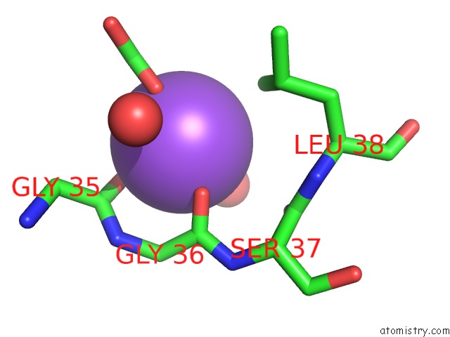
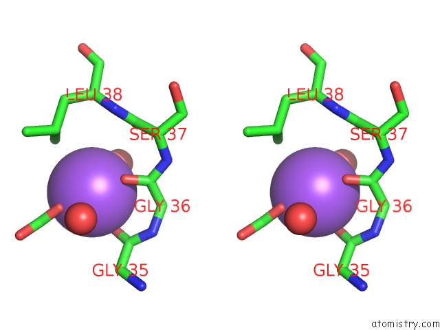
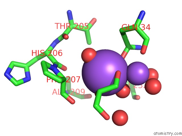
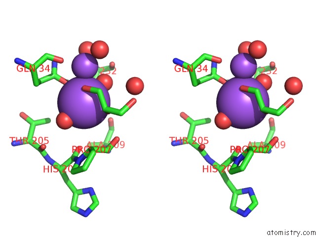
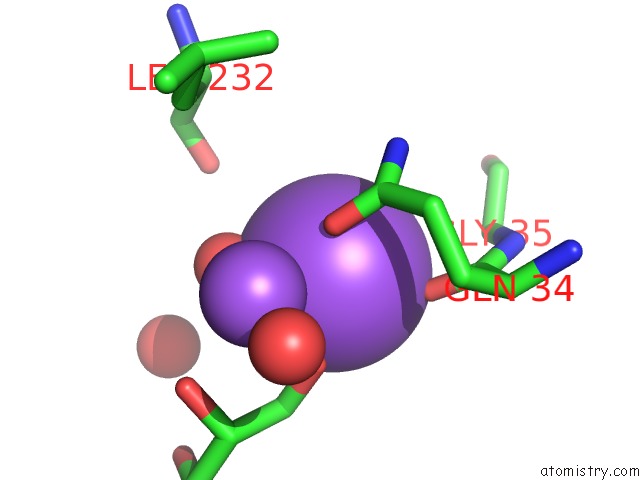
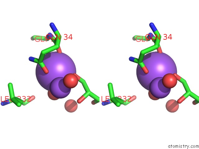
Sodium binding site 1 out of 6 in 4pmo
Go back to
Sodium binding site 1 out
of 6 in the Crystal Structure of the Mycobacterium Tuberculosis Tat-Secreted Protein RV2525C, Monoclinic Crystal Form I
Mono view
Stereo pair view
Mono view
Stereo pair view
A full contact list of Sodium with other atoms in the Na binding
site number 1 of Crystal Structure of the Mycobacterium Tuberculosis Tat-Secreted Protein RV2525C, Monoclinic Crystal Form I within 5.0Å range:
|
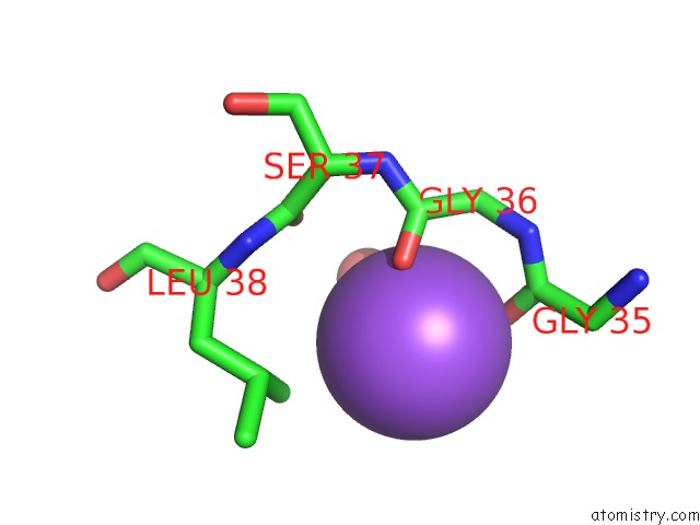
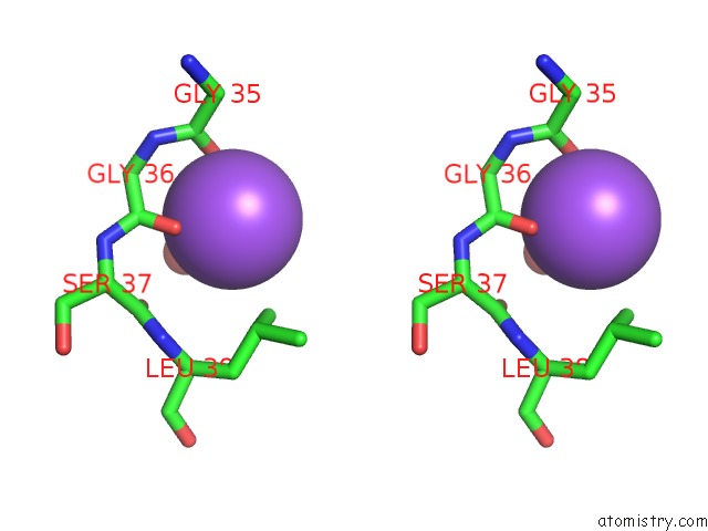
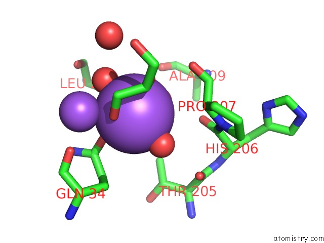
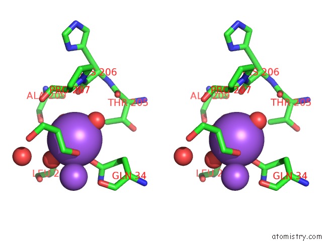
Sodium binding site 2 out of 6 in 4pmo
Go back to
Sodium binding site 2 out
of 6 in the Crystal Structure of the Mycobacterium Tuberculosis Tat-Secreted Protein RV2525C, Monoclinic Crystal Form I
Mono view
Stereo pair view
Mono view
Stereo pair view
A full contact list of Sodium with other atoms in the Na binding
site number 2 of Crystal Structure of the Mycobacterium Tuberculosis Tat-Secreted Protein RV2525C, Monoclinic Crystal Form I within 5.0Å range:
|
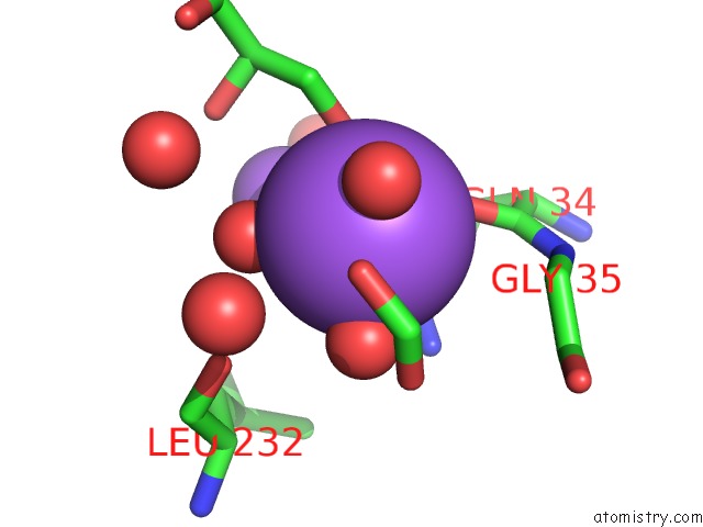
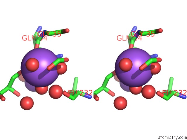
Sodium binding site 3 out of 6 in 4pmo
Go back to
Sodium binding site 3 out
of 6 in the Crystal Structure of the Mycobacterium Tuberculosis Tat-Secreted Protein RV2525C, Monoclinic Crystal Form I
Mono view
Stereo pair view
Mono view
Stereo pair view
A full contact list of Sodium with other atoms in the Na binding
site number 3 of Crystal Structure of the Mycobacterium Tuberculosis Tat-Secreted Protein RV2525C, Monoclinic Crystal Form I within 5.0Å range:
|
Sodium binding site 4 out of 6 in 4pmo
Go back to
Sodium binding site 4 out
of 6 in the Crystal Structure of the Mycobacterium Tuberculosis Tat-Secreted Protein RV2525C, Monoclinic Crystal Form I
Mono view
Stereo pair view
Mono view
Stereo pair view
A full contact list of Sodium with other atoms in the Na binding
site number 4 of Crystal Structure of the Mycobacterium Tuberculosis Tat-Secreted Protein RV2525C, Monoclinic Crystal Form I within 5.0Å range:
|
Sodium binding site 5 out of 6 in 4pmo
Go back to
Sodium binding site 5 out
of 6 in the Crystal Structure of the Mycobacterium Tuberculosis Tat-Secreted Protein RV2525C, Monoclinic Crystal Form I
Mono view
Stereo pair view
Mono view
Stereo pair view
A full contact list of Sodium with other atoms in the Na binding
site number 5 of Crystal Structure of the Mycobacterium Tuberculosis Tat-Secreted Protein RV2525C, Monoclinic Crystal Form I within 5.0Å range:
|
Sodium binding site 6 out of 6 in 4pmo
Go back to
Sodium binding site 6 out
of 6 in the Crystal Structure of the Mycobacterium Tuberculosis Tat-Secreted Protein RV2525C, Monoclinic Crystal Form I
Mono view
Stereo pair view
Mono view
Stereo pair view
A full contact list of Sodium with other atoms in the Na binding
site number 6 of Crystal Structure of the Mycobacterium Tuberculosis Tat-Secreted Protein RV2525C, Monoclinic Crystal Form I within 5.0Å range:
|
Reference:
M.Bellinzoni,
A.Haouz,
I.Miras,
S.Magnet,
G.Andre-Leroux,
R.Mukherjee,
W.Shepard,
S.T.Cole,
P.M.Alzari.
Structural Studies Suggest A Peptidoglycan Hydrolase Function For the Mycobacterium Tuberculosis Tat-Secreted Protein RV2525C. J.Struct.Biol. 2014.
ISSN: ESSN 1095-8657
PubMed: 25260828
DOI: 10.1016/J.JSB.2014.09.003
Page generated: Mon Oct 7 17:45:13 2024
ISSN: ESSN 1095-8657
PubMed: 25260828
DOI: 10.1016/J.JSB.2014.09.003
Last articles
Zn in 9MJ5Zn in 9HNW
Zn in 9G0L
Zn in 9FNE
Zn in 9DZN
Zn in 9E0I
Zn in 9D32
Zn in 9DAK
Zn in 8ZXC
Zn in 8ZUF