Sodium »
PDB 4n01-4nji »
4ni9 »
Sodium in PDB 4ni9: Crystal Structure of Human Interleukin 6 in Complex with A Modified Nucleotide Aptamer (Somamer SL1025), Form 2
Protein crystallography data
The structure of Crystal Structure of Human Interleukin 6 in Complex with A Modified Nucleotide Aptamer (Somamer SL1025), Form 2, PDB code: 4ni9
was solved by
D.Davies,
T.Edwards,
A.Gelinas,
T.Jarvis,
M.Clifton,
with X-Ray Crystallography technique. A brief refinement statistics is given in the table below:
| Resolution Low / High (Å) | 50.00 / 2.55 |
| Space group | P 32 |
| Cell size a, b, c (Å), α, β, γ (°) | 69.020, 69.020, 108.470, 90.00, 90.00, 120.00 |
| R / Rfree (%) | 18.6 / 23.9 |
Sodium Binding Sites:
The binding sites of Sodium atom in the Crystal Structure of Human Interleukin 6 in Complex with A Modified Nucleotide Aptamer (Somamer SL1025), Form 2
(pdb code 4ni9). This binding sites where shown within
5.0 Angstroms radius around Sodium atom.
In total 2 binding sites of Sodium where determined in the Crystal Structure of Human Interleukin 6 in Complex with A Modified Nucleotide Aptamer (Somamer SL1025), Form 2, PDB code: 4ni9:
Jump to Sodium binding site number: 1; 2;
In total 2 binding sites of Sodium where determined in the Crystal Structure of Human Interleukin 6 in Complex with A Modified Nucleotide Aptamer (Somamer SL1025), Form 2, PDB code: 4ni9:
Jump to Sodium binding site number: 1; 2;
Sodium binding site 1 out of 2 in 4ni9
Go back to
Sodium binding site 1 out
of 2 in the Crystal Structure of Human Interleukin 6 in Complex with A Modified Nucleotide Aptamer (Somamer SL1025), Form 2
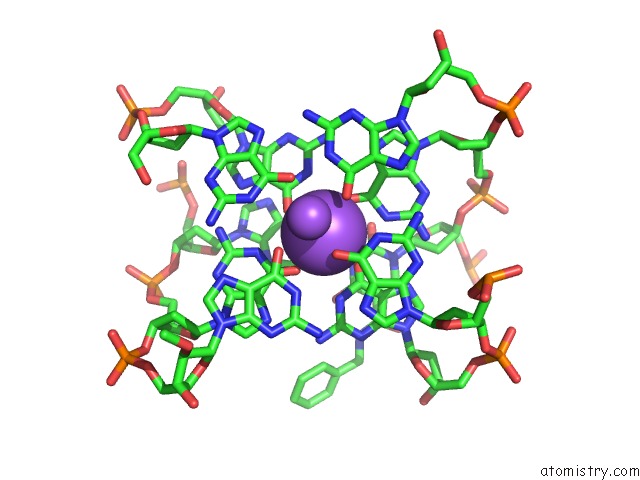
Mono view
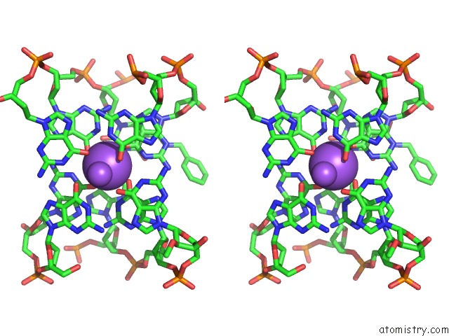
Stereo pair view
Mono view
Stereo pair view
A full contact list of Sodium with other atoms in the Na binding
site number 1 of Crystal Structure of Human Interleukin 6 in Complex with A Modified Nucleotide Aptamer (Somamer SL1025), Form 2 within 5.0Å range:
|
Sodium binding site 2 out of 2 in 4ni9
Go back to
Sodium binding site 2 out
of 2 in the Crystal Structure of Human Interleukin 6 in Complex with A Modified Nucleotide Aptamer (Somamer SL1025), Form 2
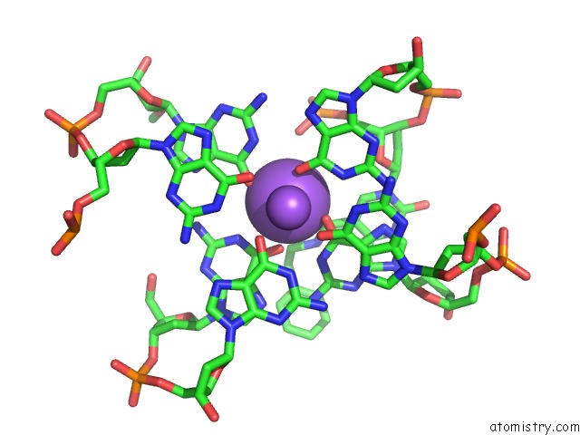
Mono view
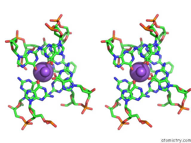
Stereo pair view
Mono view
Stereo pair view
A full contact list of Sodium with other atoms in the Na binding
site number 2 of Crystal Structure of Human Interleukin 6 in Complex with A Modified Nucleotide Aptamer (Somamer SL1025), Form 2 within 5.0Å range:
|
Reference:
A.D.Gelinas,
D.R.Davies,
T.E.Edwards,
J.C.Rohloff,
J.D.Carter,
C.Zhang,
S.Gupta,
Y.Ishikawa,
M.Hirota,
Y.Nakaishi,
T.C.Jarvis,
N.Janjic.
Crystal Structure of Interleukin-6 in Complex with A Modified Nucleic Acid Ligand. J.Biol.Chem. V. 289 8720 2014.
ISSN: ISSN 0021-9258
PubMed: 24415767
DOI: 10.1074/JBC.M113.532697
Page generated: Mon Oct 7 17:12:37 2024
ISSN: ISSN 0021-9258
PubMed: 24415767
DOI: 10.1074/JBC.M113.532697
Last articles
Zn in 9MJ5Zn in 9HNW
Zn in 9G0L
Zn in 9FNE
Zn in 9DZN
Zn in 9E0I
Zn in 9D32
Zn in 9DAK
Zn in 8ZXC
Zn in 8ZUF