Sodium »
PDB 4jvl-4khs »
4k0w »
Sodium in PDB 4k0w: X-Ray Crystal Structure of Oxa-23 A220 Duplication Clinical Variant
Enzymatic activity of X-Ray Crystal Structure of Oxa-23 A220 Duplication Clinical Variant
All present enzymatic activity of X-Ray Crystal Structure of Oxa-23 A220 Duplication Clinical Variant:
3.5.2.6;
3.5.2.6;
Protein crystallography data
The structure of X-Ray Crystal Structure of Oxa-23 A220 Duplication Clinical Variant, PDB code: 4k0w
was solved by
K.J.Kaitany,
N.V.Klinger,
D.A.Leonard,
R.A.Powers,
with X-Ray Crystallography technique. A brief refinement statistics is given in the table below:
| Resolution Low / High (Å) | 137.00 / 1.20 |
| Space group | P 21 2 21 |
| Cell size a, b, c (Å), α, β, γ (°) | 44.116, 46.662, 137.018, 90.00, 90.00, 90.00 |
| R / Rfree (%) | 18.2 / 20.2 |
Sodium Binding Sites:
The binding sites of Sodium atom in the X-Ray Crystal Structure of Oxa-23 A220 Duplication Clinical Variant
(pdb code 4k0w). This binding sites where shown within
5.0 Angstroms radius around Sodium atom.
In total 5 binding sites of Sodium where determined in the X-Ray Crystal Structure of Oxa-23 A220 Duplication Clinical Variant, PDB code: 4k0w:
Jump to Sodium binding site number: 1; 2; 3; 4; 5;
In total 5 binding sites of Sodium where determined in the X-Ray Crystal Structure of Oxa-23 A220 Duplication Clinical Variant, PDB code: 4k0w:
Jump to Sodium binding site number: 1; 2; 3; 4; 5;
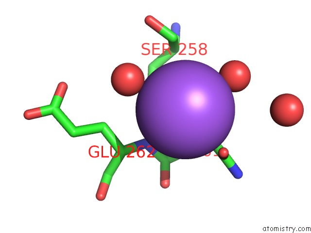
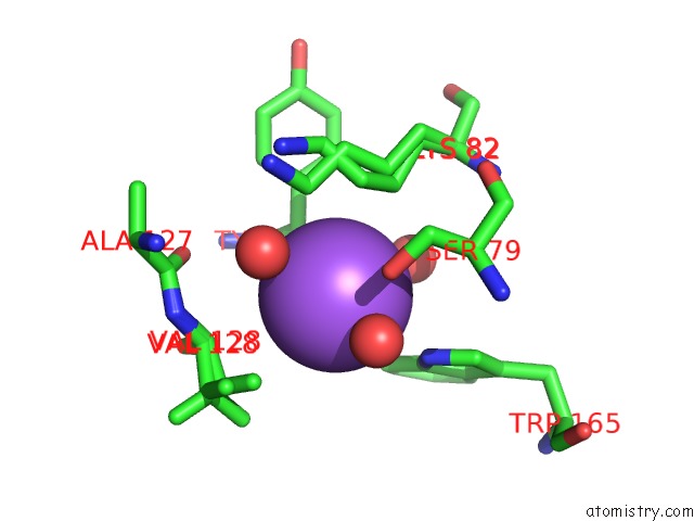
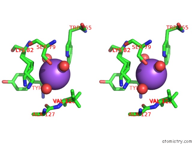
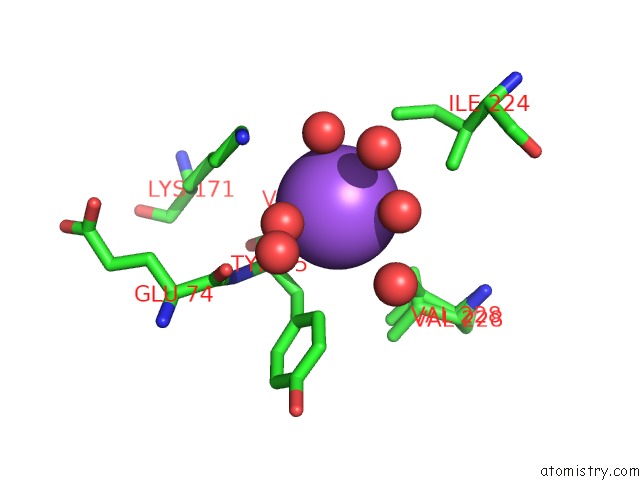
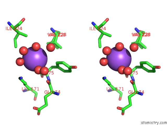
Sodium binding site 1 out of 5 in 4k0w
Go back to
Sodium binding site 1 out
of 5 in the X-Ray Crystal Structure of Oxa-23 A220 Duplication Clinical Variant
Mono view
Stereo pair view
Mono view
Stereo pair view
A full contact list of Sodium with other atoms in the Na binding
site number 1 of X-Ray Crystal Structure of Oxa-23 A220 Duplication Clinical Variant within 5.0Å range:
|
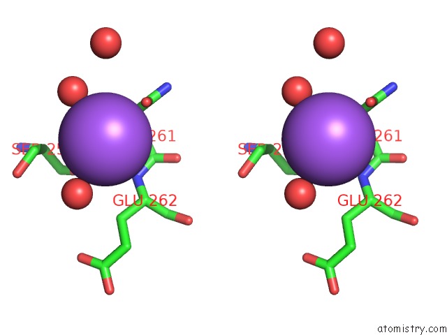
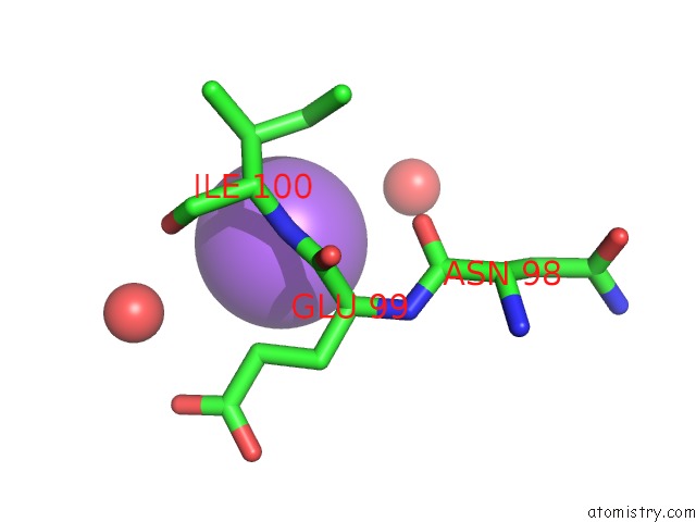
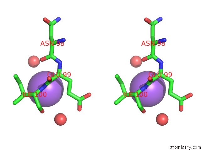
Sodium binding site 2 out of 5 in 4k0w
Go back to
Sodium binding site 2 out
of 5 in the X-Ray Crystal Structure of Oxa-23 A220 Duplication Clinical Variant
Mono view
Stereo pair view
Mono view
Stereo pair view
A full contact list of Sodium with other atoms in the Na binding
site number 2 of X-Ray Crystal Structure of Oxa-23 A220 Duplication Clinical Variant within 5.0Å range:
|
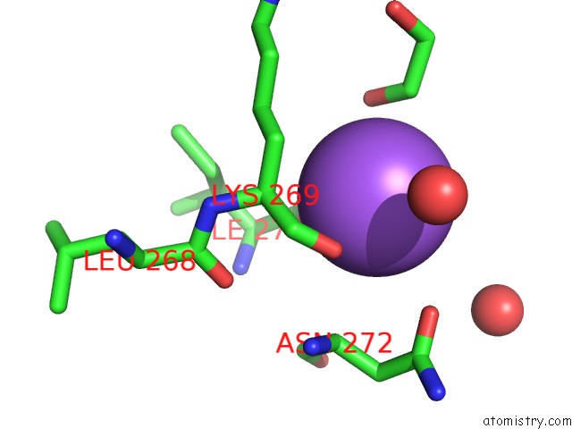
Sodium binding site 3 out of 5 in 4k0w
Go back to
Sodium binding site 3 out
of 5 in the X-Ray Crystal Structure of Oxa-23 A220 Duplication Clinical Variant
Mono view
Stereo pair view
Mono view
Stereo pair view
A full contact list of Sodium with other atoms in the Na binding
site number 3 of X-Ray Crystal Structure of Oxa-23 A220 Duplication Clinical Variant within 5.0Å range:
|
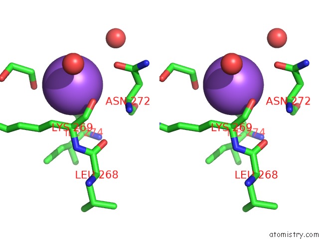
Sodium binding site 4 out of 5 in 4k0w
Go back to
Sodium binding site 4 out
of 5 in the X-Ray Crystal Structure of Oxa-23 A220 Duplication Clinical Variant
Mono view
Stereo pair view
Mono view
Stereo pair view
A full contact list of Sodium with other atoms in the Na binding
site number 4 of X-Ray Crystal Structure of Oxa-23 A220 Duplication Clinical Variant within 5.0Å range:
|
Sodium binding site 5 out of 5 in 4k0w
Go back to
Sodium binding site 5 out
of 5 in the X-Ray Crystal Structure of Oxa-23 A220 Duplication Clinical Variant
Mono view
Stereo pair view
Mono view
Stereo pair view
A full contact list of Sodium with other atoms in the Na binding
site number 5 of X-Ray Crystal Structure of Oxa-23 A220 Duplication Clinical Variant within 5.0Å range:
|
Reference:
K.C.Kaitany,
N.V.Klinger,
C.M.June,
M.E.Ramey,
R.A.Bonomo,
R.A.Powers,
D.A.Leonard.
Structures of the Class D Carbapenemases Oxa-23 and Oxa-146: Mechanistic Basis of Activity Against Carbapenems, Extended-Spectrum Cephalosporins, and Aztreonam. Antimicrob.Agents Chemother. V. 57 4848 2013.
ISSN: ISSN 0066-4804
PubMed: 23877677
DOI: 10.1128/AAC.00762-13
Page generated: Mon Oct 7 16:24:15 2024
ISSN: ISSN 0066-4804
PubMed: 23877677
DOI: 10.1128/AAC.00762-13
Last articles
Cl in 5IVRCl in 5IVQ
Cl in 5IV4
Cl in 5IV0
Cl in 5IV3
Cl in 5IUY
Cl in 5ISO
Cl in 5IS8
Cl in 5IRP
Cl in 5IUS