Sodium »
PDB 4iic-4j0n »
4ip0 »
Sodium in PDB 4ip0: X-Ray Structure of the Complex Uridine Phosphorylase From Vibrio Cholerae with Phosphate Ion at 1.29 A Resolution
Enzymatic activity of X-Ray Structure of the Complex Uridine Phosphorylase From Vibrio Cholerae with Phosphate Ion at 1.29 A Resolution
All present enzymatic activity of X-Ray Structure of the Complex Uridine Phosphorylase From Vibrio Cholerae with Phosphate Ion at 1.29 A Resolution:
2.4.2.3;
2.4.2.3;
Protein crystallography data
The structure of X-Ray Structure of the Complex Uridine Phosphorylase From Vibrio Cholerae with Phosphate Ion at 1.29 A Resolution, PDB code: 4ip0
was solved by
I.I.Prokofev,
A.A.Lashkov,
A.G.Gabdoulkhakov,
C.Betzel,
A.M.Mikhailov,
with X-Ray Crystallography technique. A brief refinement statistics is given in the table below:
| Resolution Low / High (Å) | 18.93 / 1.29 |
| Space group | P 1 |
| Cell size a, b, c (Å), α, β, γ (°) | 63.671, 71.062, 87.908, 69.63, 72.56, 85.73 |
| R / Rfree (%) | 17.2 / 20 |
Other elements in 4ip0:
The structure of X-Ray Structure of the Complex Uridine Phosphorylase From Vibrio Cholerae with Phosphate Ion at 1.29 A Resolution also contains other interesting chemical elements:
| Potassium | (K) | 1 atom |
| Chlorine | (Cl) | 2 atoms |
Sodium Binding Sites:
The binding sites of Sodium atom in the X-Ray Structure of the Complex Uridine Phosphorylase From Vibrio Cholerae with Phosphate Ion at 1.29 A Resolution
(pdb code 4ip0). This binding sites where shown within
5.0 Angstroms radius around Sodium atom.
In total 2 binding sites of Sodium where determined in the X-Ray Structure of the Complex Uridine Phosphorylase From Vibrio Cholerae with Phosphate Ion at 1.29 A Resolution, PDB code: 4ip0:
Jump to Sodium binding site number: 1; 2;
In total 2 binding sites of Sodium where determined in the X-Ray Structure of the Complex Uridine Phosphorylase From Vibrio Cholerae with Phosphate Ion at 1.29 A Resolution, PDB code: 4ip0:
Jump to Sodium binding site number: 1; 2;
Sodium binding site 1 out of 2 in 4ip0
Go back to
Sodium binding site 1 out
of 2 in the X-Ray Structure of the Complex Uridine Phosphorylase From Vibrio Cholerae with Phosphate Ion at 1.29 A Resolution
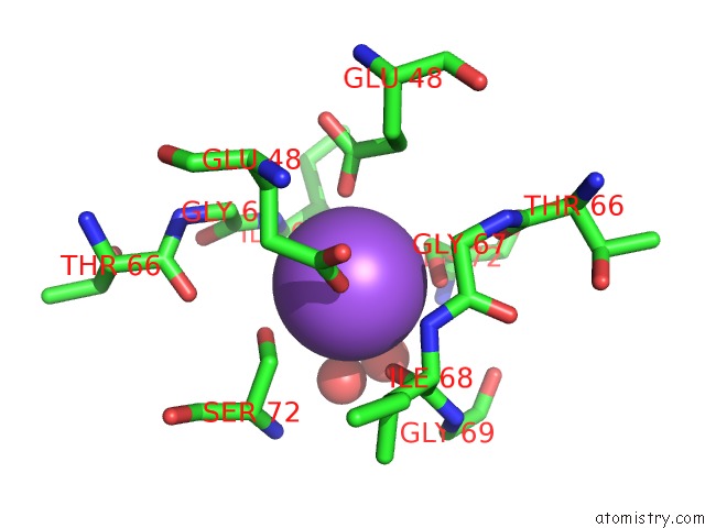
Mono view
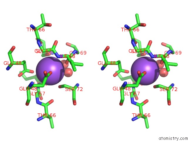
Stereo pair view
Mono view
Stereo pair view
A full contact list of Sodium with other atoms in the Na binding
site number 1 of X-Ray Structure of the Complex Uridine Phosphorylase From Vibrio Cholerae with Phosphate Ion at 1.29 A Resolution within 5.0Å range:
|
Sodium binding site 2 out of 2 in 4ip0
Go back to
Sodium binding site 2 out
of 2 in the X-Ray Structure of the Complex Uridine Phosphorylase From Vibrio Cholerae with Phosphate Ion at 1.29 A Resolution
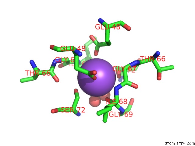
Mono view
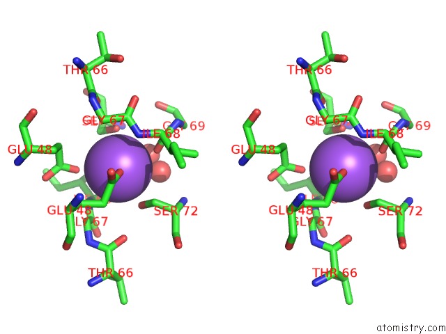
Stereo pair view
Mono view
Stereo pair view
A full contact list of Sodium with other atoms in the Na binding
site number 2 of X-Ray Structure of the Complex Uridine Phosphorylase From Vibrio Cholerae with Phosphate Ion at 1.29 A Resolution within 5.0Å range:
|
Reference:
I.I.Prokofev,
A.A.Lashkov,
A.G.Gabdoulkhakov,
C.Betzel,
A.M.Mikhailov.
X-Ray Structure of the Complex Uridine Phosphorylase From Vibrio Cholerae with Phosphate Ion at 1.29 A Resolution To Be Published.
Page generated: Sun Aug 17 19:53:13 2025
Last articles
Na in 6O48Na in 6O3C
Na in 6O3A
Na in 6NZ8
Na in 6O08
Na in 6NLH
Na in 6NYP
Na in 6NXS
Na in 6NXX
Na in 6NXY