Sodium »
PDB 4iic-4j0n »
4imk »
Sodium in PDB 4imk: Uncrossed Fab Binding to Human Angiopoietin 2
Protein crystallography data
The structure of Uncrossed Fab Binding to Human Angiopoietin 2, PDB code: 4imk
was solved by
S.Fenn,
C.Schiller,
J.J.Griese,
K.-P.Hopfner,
H.Kettenberger,
with X-Ray Crystallography technique. A brief refinement statistics is given in the table below:
| Resolution Low / High (Å) | 46.76 / 2.20 |
| Space group | P 21 21 21 |
| Cell size a, b, c (Å), α, β, γ (°) | 65.969, 86.670, 205.813, 90.00, 90.00, 90.00 |
| R / Rfree (%) | 19.9 / 22.6 |
Sodium Binding Sites:
The binding sites of Sodium atom in the Uncrossed Fab Binding to Human Angiopoietin 2
(pdb code 4imk). This binding sites where shown within
5.0 Angstroms radius around Sodium atom.
In total only one binding site of Sodium was determined in the Uncrossed Fab Binding to Human Angiopoietin 2, PDB code: 4imk:
In total only one binding site of Sodium was determined in the Uncrossed Fab Binding to Human Angiopoietin 2, PDB code: 4imk:
Sodium binding site 1 out of 1 in 4imk
Go back to
Sodium binding site 1 out
of 1 in the Uncrossed Fab Binding to Human Angiopoietin 2
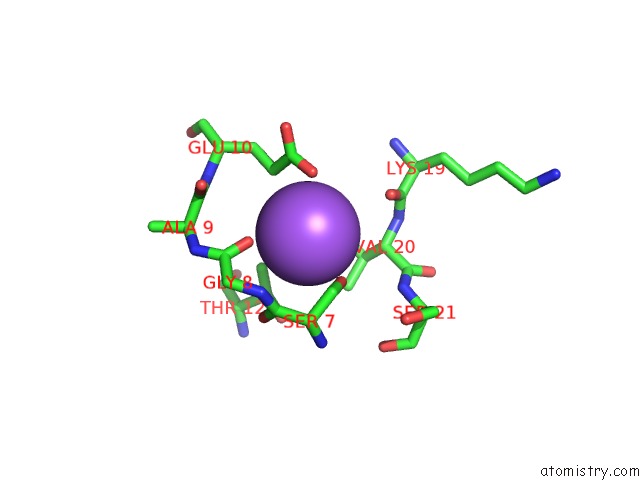
Mono view
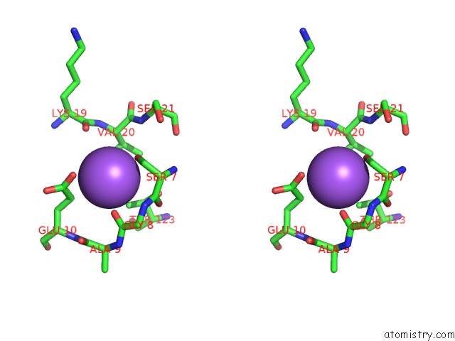
Stereo pair view
Mono view
Stereo pair view
A full contact list of Sodium with other atoms in the Na binding
site number 1 of Uncrossed Fab Binding to Human Angiopoietin 2 within 5.0Å range:
|
Reference:
S.Fenn,
C.B.Schiller,
J.J.Griese,
H.Duerr,
S.Imhof-Jung,
C.Gassner,
J.Moelleken,
J.T.Regula,
W.Schaefer,
M.Thomas,
C.Klein,
K.P.Hopfner,
H.Kettenberger.
Crystal Structure of An Anti-ANG2 Crossfab Demonstrates Complete Structural and Functional Integrity of the Variable Domain. Plos One V. 8 61953 2013.
ISSN: ESSN 1932-6203
PubMed: 23613981
DOI: 10.1371/JOURNAL.PONE.0061953
Page generated: Mon Oct 7 16:03:17 2024
ISSN: ESSN 1932-6203
PubMed: 23613981
DOI: 10.1371/JOURNAL.PONE.0061953
Last articles
Zn in 9J0NZn in 9J0O
Zn in 9J0P
Zn in 9FJX
Zn in 9EKB
Zn in 9C0F
Zn in 9CAH
Zn in 9CH0
Zn in 9CH3
Zn in 9CH1