Sodium »
PDB 4fpa-4gfi »
4gbj »
Sodium in PDB 4gbj: Crystal Structure of Nad-Binding 6-Phosphogluconate Dehydrogenase From Dyadobacter Fermentans
Protein crystallography data
The structure of Crystal Structure of Nad-Binding 6-Phosphogluconate Dehydrogenase From Dyadobacter Fermentans, PDB code: 4gbj
was solved by
K.Michalska,
J.Holowicki,
S.Clancy,
A.Joachimiak,
Midwest Center Forstructural Genomics (Mcsg),
with X-Ray Crystallography technique. A brief refinement statistics is given in the table below:
| Resolution Low / High (Å) | 30.01 / 2.05 |
| Space group | P 21 21 21 |
| Cell size a, b, c (Å), α, β, γ (°) | 79.633, 89.645, 151.506, 90.00, 90.00, 90.00 |
| R / Rfree (%) | 16 / 19.7 |
Sodium Binding Sites:
The binding sites of Sodium atom in the Crystal Structure of Nad-Binding 6-Phosphogluconate Dehydrogenase From Dyadobacter Fermentans
(pdb code 4gbj). This binding sites where shown within
5.0 Angstroms radius around Sodium atom.
In total 4 binding sites of Sodium where determined in the Crystal Structure of Nad-Binding 6-Phosphogluconate Dehydrogenase From Dyadobacter Fermentans, PDB code: 4gbj:
Jump to Sodium binding site number: 1; 2; 3; 4;
In total 4 binding sites of Sodium where determined in the Crystal Structure of Nad-Binding 6-Phosphogluconate Dehydrogenase From Dyadobacter Fermentans, PDB code: 4gbj:
Jump to Sodium binding site number: 1; 2; 3; 4;
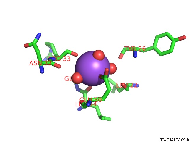
Sodium binding site 1 out of 4 in 4gbj
Go back to
Sodium binding site 1 out
of 4 in the Crystal Structure of Nad-Binding 6-Phosphogluconate Dehydrogenase From Dyadobacter Fermentans
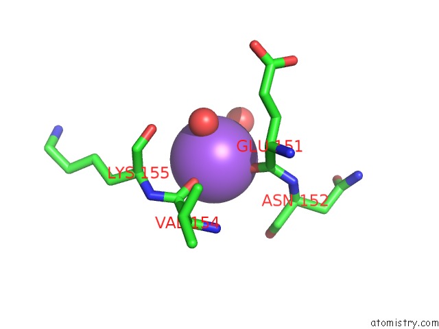
Mono view
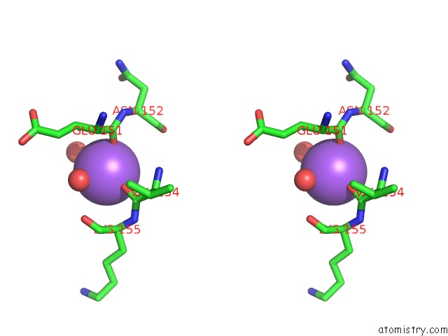
Stereo pair view
Mono view
Stereo pair view
A full contact list of Sodium with other atoms in the Na binding
site number 1 of Crystal Structure of Nad-Binding 6-Phosphogluconate Dehydrogenase From Dyadobacter Fermentans within 5.0Å range:
|
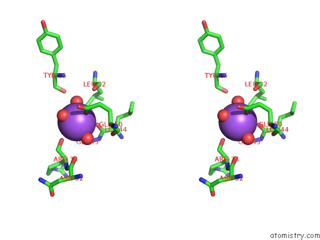
Sodium binding site 2 out of 4 in 4gbj
Go back to
Sodium binding site 2 out
of 4 in the Crystal Structure of Nad-Binding 6-Phosphogluconate Dehydrogenase From Dyadobacter Fermentans
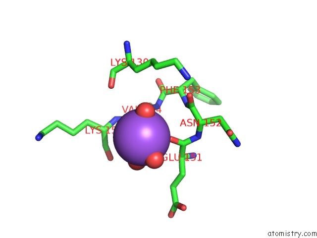
Mono view
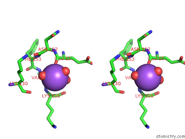
Stereo pair view
Mono view
Stereo pair view
A full contact list of Sodium with other atoms in the Na binding
site number 2 of Crystal Structure of Nad-Binding 6-Phosphogluconate Dehydrogenase From Dyadobacter Fermentans within 5.0Å range:
|
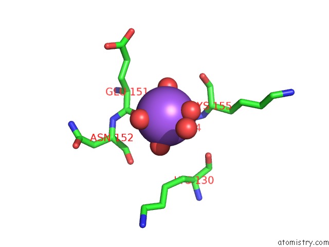
Sodium binding site 3 out of 4 in 4gbj
Go back to
Sodium binding site 3 out
of 4 in the Crystal Structure of Nad-Binding 6-Phosphogluconate Dehydrogenase From Dyadobacter Fermentans
Mono view
Stereo pair view
Mono view
Stereo pair view
A full contact list of Sodium with other atoms in the Na binding
site number 3 of Crystal Structure of Nad-Binding 6-Phosphogluconate Dehydrogenase From Dyadobacter Fermentans within 5.0Å range:
|
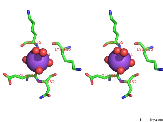
Sodium binding site 4 out of 4 in 4gbj
Go back to
Sodium binding site 4 out
of 4 in the Crystal Structure of Nad-Binding 6-Phosphogluconate Dehydrogenase From Dyadobacter Fermentans
Mono view
Stereo pair view
Mono view
Stereo pair view
A full contact list of Sodium with other atoms in the Na binding
site number 4 of Crystal Structure of Nad-Binding 6-Phosphogluconate Dehydrogenase From Dyadobacter Fermentans within 5.0Å range:
|
Reference:
K.Michalska,
J.Holowicki,
S.Clancy,
A.Joachimiak,
Midwest Center For Structural Genomics (Mcsg).
Crystal Structure of Nad-Binding 6-Phosphogluconate Dehydrogenase From Dyadobacter Fermentans To Be Published.
Page generated: Mon Oct 7 15:32:23 2024
Last articles
Ca in 5NMRCa in 5NN9
Ca in 5NM8
Ca in 5NH8
Ca in 5NL7
Ca in 5NIN
Ca in 5NGQ
Ca in 5NH5
Ca in 5NGY
Ca in 5NG1