Sodium »
PDB 4fpa-4gfi »
4fs2 »
Sodium in PDB 4fs2: Base Pairing Mechanism of N2,3-Ethenoguanine with Dctp By Human Polymerase Iota
Enzymatic activity of Base Pairing Mechanism of N2,3-Ethenoguanine with Dctp By Human Polymerase Iota
All present enzymatic activity of Base Pairing Mechanism of N2,3-Ethenoguanine with Dctp By Human Polymerase Iota:
2.7.7.7;
2.7.7.7;
Protein crystallography data
The structure of Base Pairing Mechanism of N2,3-Ethenoguanine with Dctp By Human Polymerase Iota, PDB code: 4fs2
was solved by
L.Zhao,
with X-Ray Crystallography technique. A brief refinement statistics is given in the table below:
| Resolution Low / High (Å) | 84.26 / 2.05 |
| Space group | P 65 2 2 |
| Cell size a, b, c (Å), α, β, γ (°) | 97.298, 97.298, 202.906, 90.00, 90.00, 120.00 |
| R / Rfree (%) | 21.4 / 26.1 |
Other elements in 4fs2:
The structure of Base Pairing Mechanism of N2,3-Ethenoguanine with Dctp By Human Polymerase Iota also contains other interesting chemical elements:
| Fluorine | (F) | 1 atom |
| Magnesium | (Mg) | 3 atoms |
Sodium Binding Sites:
The binding sites of Sodium atom in the Base Pairing Mechanism of N2,3-Ethenoguanine with Dctp By Human Polymerase Iota
(pdb code 4fs2). This binding sites where shown within
5.0 Angstroms radius around Sodium atom.
In total only one binding site of Sodium was determined in the Base Pairing Mechanism of N2,3-Ethenoguanine with Dctp By Human Polymerase Iota, PDB code: 4fs2:
In total only one binding site of Sodium was determined in the Base Pairing Mechanism of N2,3-Ethenoguanine with Dctp By Human Polymerase Iota, PDB code: 4fs2:
Sodium binding site 1 out of 1 in 4fs2
Go back to
Sodium binding site 1 out
of 1 in the Base Pairing Mechanism of N2,3-Ethenoguanine with Dctp By Human Polymerase Iota
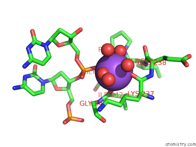
Mono view
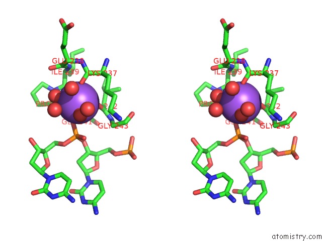
Stereo pair view
Mono view
Stereo pair view
A full contact list of Sodium with other atoms in the Na binding
site number 1 of Base Pairing Mechanism of N2,3-Ethenoguanine with Dctp By Human Polymerase Iota within 5.0Å range:
|
Reference:
L.Zhao,
M.G.Pence,
P.P.Christov,
Z.Wawrzak,
J.Y.Choi,
C.J.Rizzo,
M.Egli,
F.P.Guengerich.
Basis of Miscoding of the Dna Adduct N2,3-Ethenoguanine By Human Y-Family Dna Polymerases. J.Biol.Chem. V. 287 35516 2012.
ISSN: ISSN 0021-9258
PubMed: 22910910
DOI: 10.1074/JBC.M112.403253
Page generated: Mon Oct 7 15:27:30 2024
ISSN: ISSN 0021-9258
PubMed: 22910910
DOI: 10.1074/JBC.M112.403253
Last articles
Zn in 9J0NZn in 9J0O
Zn in 9J0P
Zn in 9FJX
Zn in 9EKB
Zn in 9C0F
Zn in 9CAH
Zn in 9CH0
Zn in 9CH3
Zn in 9CH1