Sodium »
PDB 4dy7-4egs »
4e3q »
Sodium in PDB 4e3q: Pmp-Bound Form of Aminotransferase Crystal Structure From Vibrio Fluvialis
Protein crystallography data
The structure of Pmp-Bound Form of Aminotransferase Crystal Structure From Vibrio Fluvialis, PDB code: 4e3q
was solved by
K.S.Midelfort,
R.Kumar,
S.Han,
M.J.Karmilowicz,
K.Mcconnell,
D.K.Gehlhaar,
A.Mistry,
J.S.Chang,
M.Anderson,
A.Vilalobos,
J.Minshull,
S.Govindarajan,
J.W.Wong,
with X-Ray Crystallography technique. A brief refinement statistics is given in the table below:
| Resolution Low / High (Å) | 49.79 / 1.90 |
| Space group | P 21 21 21 |
| Cell size a, b, c (Å), α, β, γ (°) | 63.071, 162.182, 180.398, 90.00, 90.00, 90.00 |
| R / Rfree (%) | 17.4 / 21 |
Sodium Binding Sites:
The binding sites of Sodium atom in the Pmp-Bound Form of Aminotransferase Crystal Structure From Vibrio Fluvialis
(pdb code 4e3q). This binding sites where shown within
5.0 Angstroms radius around Sodium atom.
In total 4 binding sites of Sodium where determined in the Pmp-Bound Form of Aminotransferase Crystal Structure From Vibrio Fluvialis, PDB code: 4e3q:
Jump to Sodium binding site number: 1; 2; 3; 4;
In total 4 binding sites of Sodium where determined in the Pmp-Bound Form of Aminotransferase Crystal Structure From Vibrio Fluvialis, PDB code: 4e3q:
Jump to Sodium binding site number: 1; 2; 3; 4;
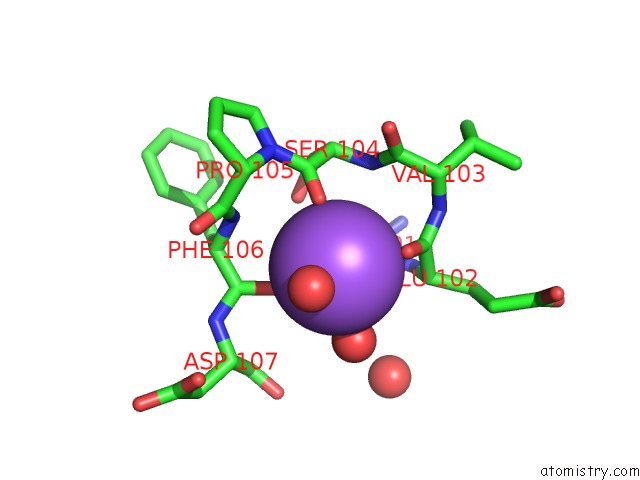
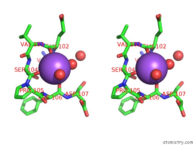
Sodium binding site 1 out of 4 in 4e3q
Go back to
Sodium binding site 1 out
of 4 in the Pmp-Bound Form of Aminotransferase Crystal Structure From Vibrio Fluvialis
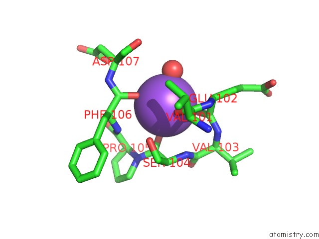
Mono view
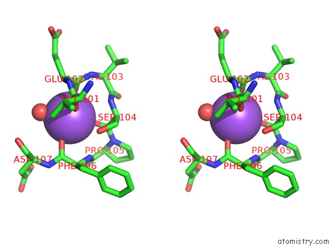
Stereo pair view
Mono view
Stereo pair view
A full contact list of Sodium with other atoms in the Na binding
site number 1 of Pmp-Bound Form of Aminotransferase Crystal Structure From Vibrio Fluvialis within 5.0Å range:
|
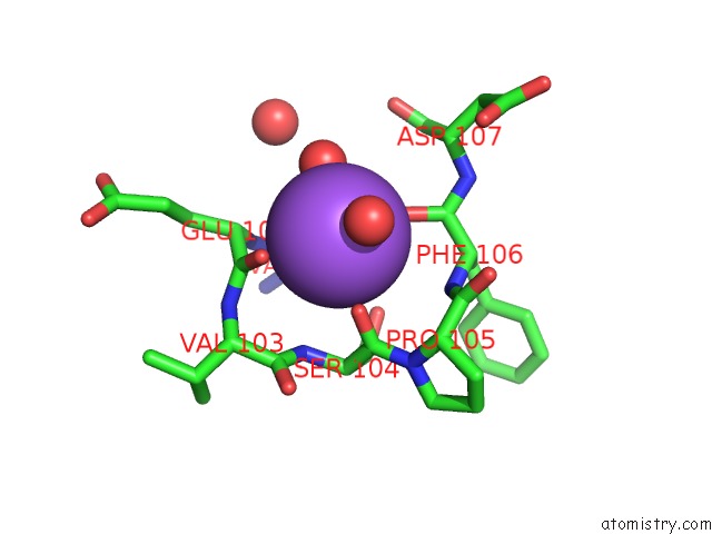
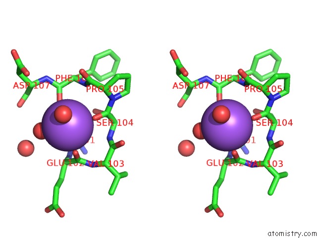
Sodium binding site 2 out of 4 in 4e3q
Go back to
Sodium binding site 2 out
of 4 in the Pmp-Bound Form of Aminotransferase Crystal Structure From Vibrio Fluvialis
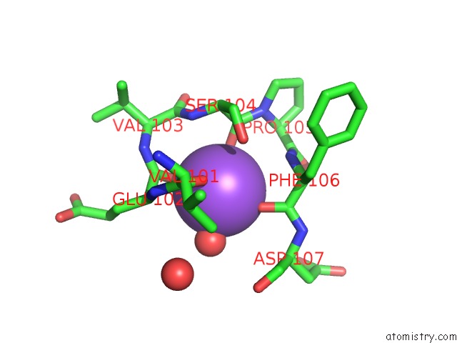
Mono view
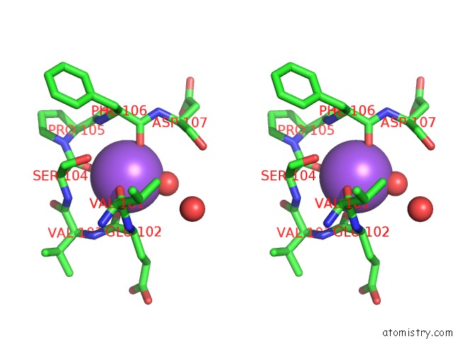
Stereo pair view
Mono view
Stereo pair view
A full contact list of Sodium with other atoms in the Na binding
site number 2 of Pmp-Bound Form of Aminotransferase Crystal Structure From Vibrio Fluvialis within 5.0Å range:
|
Sodium binding site 3 out of 4 in 4e3q
Go back to
Sodium binding site 3 out
of 4 in the Pmp-Bound Form of Aminotransferase Crystal Structure From Vibrio Fluvialis
Mono view
Stereo pair view
Mono view
Stereo pair view
A full contact list of Sodium with other atoms in the Na binding
site number 3 of Pmp-Bound Form of Aminotransferase Crystal Structure From Vibrio Fluvialis within 5.0Å range:
|
Sodium binding site 4 out of 4 in 4e3q
Go back to
Sodium binding site 4 out
of 4 in the Pmp-Bound Form of Aminotransferase Crystal Structure From Vibrio Fluvialis
Mono view
Stereo pair view
Mono view
Stereo pair view
A full contact list of Sodium with other atoms in the Na binding
site number 4 of Pmp-Bound Form of Aminotransferase Crystal Structure From Vibrio Fluvialis within 5.0Å range:
|
Reference:
K.S.Midelfort,
R.Kumar,
S.Han,
M.J.Karmilowicz,
K.Mcconnell,
D.K.Gehlhaar,
A.Mistry,
J.S.Chang,
M.Anderson,
A.Villalobos,
J.Minshull,
S.Govindarajan,
J.W.Wong.
Redesigning and Characterizing the Substrate Specificity and Activity of Vibrio Fluvialis Aminotransferase For the Synthesis of Imagabalin. Protein Eng.Des.Sel. V. 26 25 2013.
ISSN: ISSN 1741-0126
PubMed: 23012440
DOI: 10.1093/PROTEIN/GZS065
Page generated: Mon Oct 7 15:05:53 2024
ISSN: ISSN 1741-0126
PubMed: 23012440
DOI: 10.1093/PROTEIN/GZS065
Last articles
Zn in 9J0NZn in 9J0O
Zn in 9J0P
Zn in 9FJX
Zn in 9EKB
Zn in 9C0F
Zn in 9CAH
Zn in 9CH0
Zn in 9CH3
Zn in 9CH1