Sodium »
PDB 4dy7-4egs »
4e11 »
Sodium in PDB 4e11: Crystal Structure of Kynurenine Formamidase From Drosophila Melanogaster
Enzymatic activity of Crystal Structure of Kynurenine Formamidase From Drosophila Melanogaster
All present enzymatic activity of Crystal Structure of Kynurenine Formamidase From Drosophila Melanogaster:
3.5.1.9;
3.5.1.9;
Protein crystallography data
The structure of Crystal Structure of Kynurenine Formamidase From Drosophila Melanogaster, PDB code: 4e11
was solved by
Q.Han,
H.Robinson,
J.Li,
with X-Ray Crystallography technique. A brief refinement statistics is given in the table below:
| Resolution Low / High (Å) | 42.84 / 2.00 |
| Space group | C 1 2 1 |
| Cell size a, b, c (Å), α, β, γ (°) | 75.771, 47.085, 85.683, 90.00, 90.34, 90.00 |
| R / Rfree (%) | 22.5 / 27.3 |
Sodium Binding Sites:
The binding sites of Sodium atom in the Crystal Structure of Kynurenine Formamidase From Drosophila Melanogaster
(pdb code 4e11). This binding sites where shown within
5.0 Angstroms radius around Sodium atom.
In total only one binding site of Sodium was determined in the Crystal Structure of Kynurenine Formamidase From Drosophila Melanogaster, PDB code: 4e11:
In total only one binding site of Sodium was determined in the Crystal Structure of Kynurenine Formamidase From Drosophila Melanogaster, PDB code: 4e11:
Sodium binding site 1 out of 1 in 4e11
Go back to
Sodium binding site 1 out
of 1 in the Crystal Structure of Kynurenine Formamidase From Drosophila Melanogaster
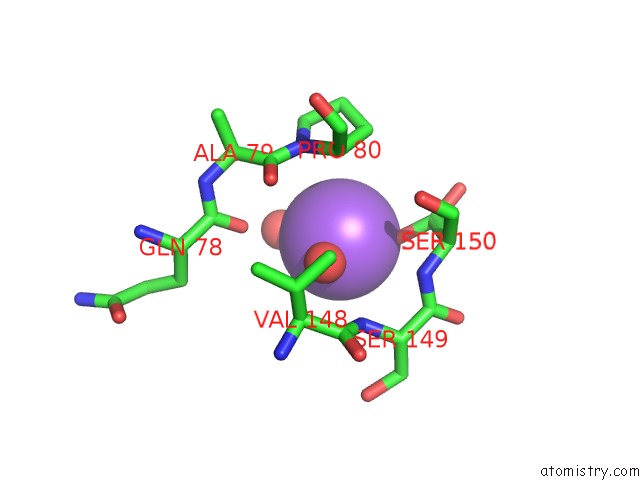
Mono view
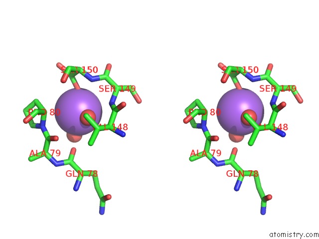
Stereo pair view
Mono view
Stereo pair view
A full contact list of Sodium with other atoms in the Na binding
site number 1 of Crystal Structure of Kynurenine Formamidase From Drosophila Melanogaster within 5.0Å range:
|
Reference:
Q.Han,
H.Robinson,
J.Li.
Biochemical Identification and Crystal Structure of Kynurenine Formamidase From Drosophila Melanogaster. Biochem.J. V. 446 253 2012.
ISSN: ISSN 0264-6021
PubMed: 22690733
DOI: 10.1042/BJ20120416
Page generated: Mon Oct 7 15:05:33 2024
ISSN: ISSN 0264-6021
PubMed: 22690733
DOI: 10.1042/BJ20120416
Last articles
Zn in 9J0NZn in 9J0O
Zn in 9J0P
Zn in 9FJX
Zn in 9EKB
Zn in 9C0F
Zn in 9CAH
Zn in 9CH0
Zn in 9CH3
Zn in 9CH1