Sodium »
PDB 4dy7-4egs »
4e0k »
Sodium in PDB 4e0k: Crystal Structure of the Amyloid-Fibril Forming Peptide Kdwsfy Derived From Human Beta 2 Microglobulin (58-63)
Protein crystallography data
The structure of Crystal Structure of the Amyloid-Fibril Forming Peptide Kdwsfy Derived From Human Beta 2 Microglobulin (58-63), PDB code: 4e0k
was solved by
M.Zhao,
C.Liu,
M.R.Sawaya,
D.Eisenberg,
with X-Ray Crystallography technique. A brief refinement statistics is given in the table below:
| Resolution Low / High (Å) | 14.98 / 0.97 |
| Space group | P 61 |
| Cell size a, b, c (Å), α, β, γ (°) | 62.370, 62.370, 11.730, 90.00, 90.00, 120.00 |
| R / Rfree (%) | 11.2 / 13.1 |
Sodium Binding Sites:
The binding sites of Sodium atom in the Crystal Structure of the Amyloid-Fibril Forming Peptide Kdwsfy Derived From Human Beta 2 Microglobulin (58-63)
(pdb code 4e0k). This binding sites where shown within
5.0 Angstroms radius around Sodium atom.
In total only one binding site of Sodium was determined in the Crystal Structure of the Amyloid-Fibril Forming Peptide Kdwsfy Derived From Human Beta 2 Microglobulin (58-63), PDB code: 4e0k:
In total only one binding site of Sodium was determined in the Crystal Structure of the Amyloid-Fibril Forming Peptide Kdwsfy Derived From Human Beta 2 Microglobulin (58-63), PDB code: 4e0k:
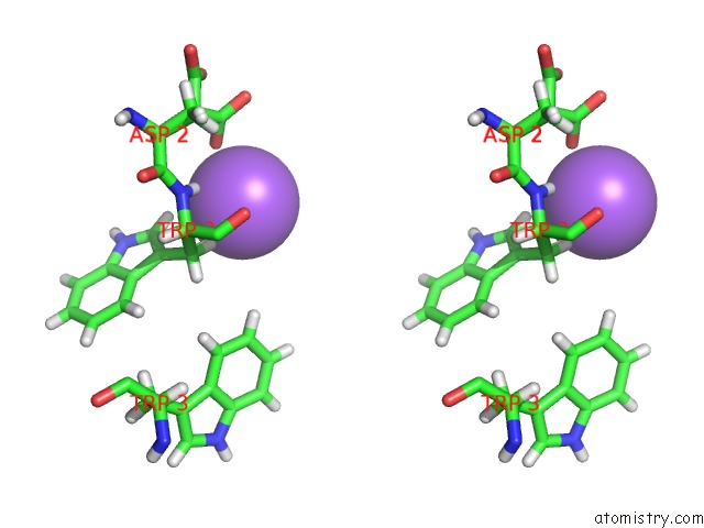
Sodium binding site 1 out of 1 in 4e0k
Go back to
Sodium binding site 1 out
of 1 in the Crystal Structure of the Amyloid-Fibril Forming Peptide Kdwsfy Derived From Human Beta 2 Microglobulin (58-63)
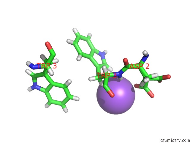
Mono view
Stereo pair view
Mono view
Stereo pair view
A full contact list of Sodium with other atoms in the Na binding
site number 1 of Crystal Structure of the Amyloid-Fibril Forming Peptide Kdwsfy Derived From Human Beta 2 Microglobulin (58-63) within 5.0Å range:
|
Reference:
C.Liu,
M.Zhao,
L.Jiang,
P.N.Cheng,
J.Park,
M.R.Sawaya,
A.Pensalfini,
D.Gou,
A.J.Berk,
C.G.Glabe,
J.Nowick,
D.Eisenberg.
Out-of-Register Beta-Sheets Suggest A Pathway to Toxic Amyloid Aggregates. Proc.Natl.Acad.Sci.Usa V. 109 20913 2012.
ISSN: ISSN 0027-8424
PubMed: 23213214
DOI: 10.1073/PNAS.1218792109
Page generated: Mon Oct 7 15:05:11 2024
ISSN: ISSN 0027-8424
PubMed: 23213214
DOI: 10.1073/PNAS.1218792109
Last articles
Zn in 9J0NZn in 9J0O
Zn in 9J0P
Zn in 9FJX
Zn in 9EKB
Zn in 9C0F
Zn in 9CAH
Zn in 9CH0
Zn in 9CH3
Zn in 9CH1