Sodium »
PDB 4def-4dxz »
4dxz »
Sodium in PDB 4dxz: Crystal Structure of A Plig-Ec Mutant, A Periplasmic Lysozyme Inhibitor of G-Type Lysozyme From Escherichia Coli
Protein crystallography data
The structure of Crystal Structure of A Plig-Ec Mutant, A Periplasmic Lysozyme Inhibitor of G-Type Lysozyme From Escherichia Coli, PDB code: 4dxz
was solved by
S.Leysen,
S.Vanheuverzwijn,
K.Van Asten,
L.Vanderkelen,
C.W.Michiels,
S.V.Strelkov,
with X-Ray Crystallography technique. A brief refinement statistics is given in the table below:
| Resolution Low / High (Å) | 40.16 / 1.25 |
| Space group | I 41 |
| Cell size a, b, c (Å), α, β, γ (°) | 80.316, 80.316, 31.098, 90.00, 90.00, 90.00 |
| R / Rfree (%) | 14.1 / 17.1 |
Sodium Binding Sites:
The binding sites of Sodium atom in the Crystal Structure of A Plig-Ec Mutant, A Periplasmic Lysozyme Inhibitor of G-Type Lysozyme From Escherichia Coli
(pdb code 4dxz). This binding sites where shown within
5.0 Angstroms radius around Sodium atom.
In total 3 binding sites of Sodium where determined in the Crystal Structure of A Plig-Ec Mutant, A Periplasmic Lysozyme Inhibitor of G-Type Lysozyme From Escherichia Coli, PDB code: 4dxz:
Jump to Sodium binding site number: 1; 2; 3;
In total 3 binding sites of Sodium where determined in the Crystal Structure of A Plig-Ec Mutant, A Periplasmic Lysozyme Inhibitor of G-Type Lysozyme From Escherichia Coli, PDB code: 4dxz:
Jump to Sodium binding site number: 1; 2; 3;
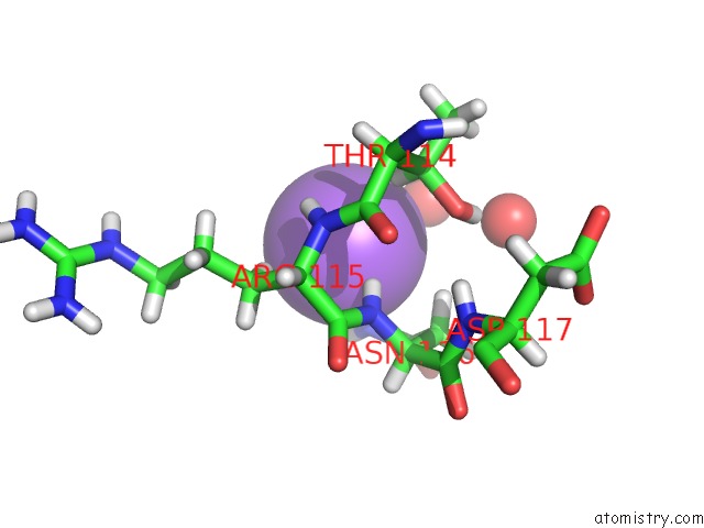
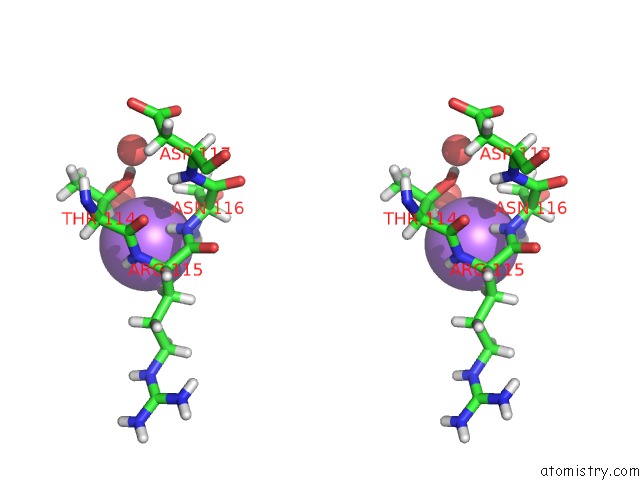
Sodium binding site 1 out of 3 in 4dxz
Go back to
Sodium binding site 1 out
of 3 in the Crystal Structure of A Plig-Ec Mutant, A Periplasmic Lysozyme Inhibitor of G-Type Lysozyme From Escherichia Coli
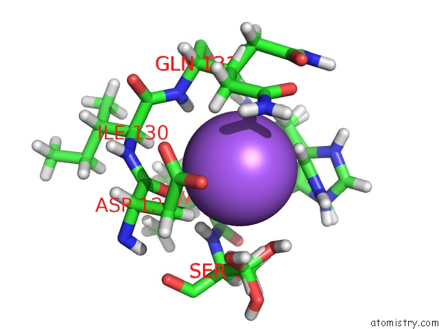
Mono view
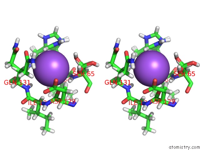
Stereo pair view
Mono view
Stereo pair view
A full contact list of Sodium with other atoms in the Na binding
site number 1 of Crystal Structure of A Plig-Ec Mutant, A Periplasmic Lysozyme Inhibitor of G-Type Lysozyme From Escherichia Coli within 5.0Å range:
|
Sodium binding site 2 out of 3 in 4dxz
Go back to
Sodium binding site 2 out
of 3 in the Crystal Structure of A Plig-Ec Mutant, A Periplasmic Lysozyme Inhibitor of G-Type Lysozyme From Escherichia Coli
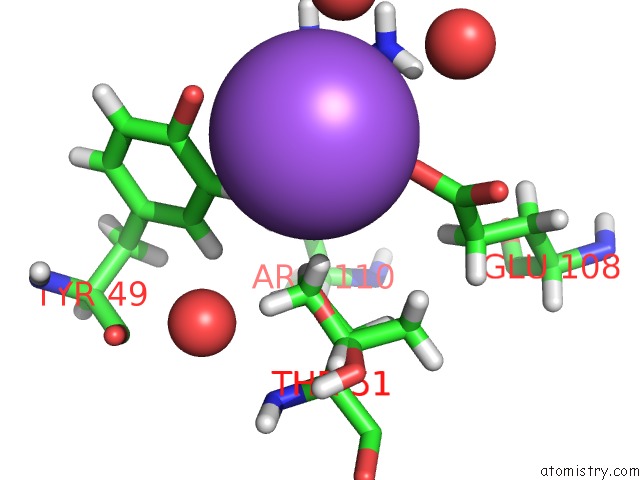
Mono view
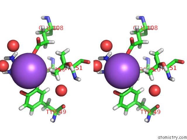
Stereo pair view
Mono view
Stereo pair view
A full contact list of Sodium with other atoms in the Na binding
site number 2 of Crystal Structure of A Plig-Ec Mutant, A Periplasmic Lysozyme Inhibitor of G-Type Lysozyme From Escherichia Coli within 5.0Å range:
|
Sodium binding site 3 out of 3 in 4dxz
Go back to
Sodium binding site 3 out
of 3 in the Crystal Structure of A Plig-Ec Mutant, A Periplasmic Lysozyme Inhibitor of G-Type Lysozyme From Escherichia Coli
Mono view
Stereo pair view
Mono view
Stereo pair view
A full contact list of Sodium with other atoms in the Na binding
site number 3 of Crystal Structure of A Plig-Ec Mutant, A Periplasmic Lysozyme Inhibitor of G-Type Lysozyme From Escherichia Coli within 5.0Å range:
|
Reference:
S.Leysen,
L.Vandekelen,
C.W.Michiels,
S.V.Strelkov.
Crystal Structure of A Plig-Ec Mutant, A Periplasmic Lysozyme Inhibitor of G-Type Lysozyme From Escherichia Coli To Be Published.
Page generated: Mon Oct 7 15:01:43 2024
Last articles
F in 4EWQF in 4EQU
F in 4EST
F in 4ENH
F in 4EPX
F in 4ENC
F in 4ENB
F in 4EMV
F in 4ENA
F in 4EN5