Sodium »
PDB 4def-4dxz »
4dxk »
Sodium in PDB 4dxk: Crystal Structure of An Enolase (Mandelate Racemase Subgroup, Target Efi-502086) From Agrobacterium Tumefaciens, with A Succinimide Residue, Na and Phosphate
Protein crystallography data
The structure of Crystal Structure of An Enolase (Mandelate Racemase Subgroup, Target Efi-502086) From Agrobacterium Tumefaciens, with A Succinimide Residue, Na and Phosphate, PDB code: 4dxk
was solved by
M.W.Vetting,
J.T.Bouvier,
R.Toro,
R.Bhosle,
N.F.Al Obaidi,
L.L.Morisco,
S.R.Wasserman,
S.Sojitra,
H.J.Imker,
J.A.Gerlt,
S.C.Almo,
Enzyme Functioninitiative (Efi),
with X-Ray Crystallography technique. A brief refinement statistics is given in the table below:
| Resolution Low / High (Å) | 20.46 / 1.25 |
| Space group | I 4 2 2 |
| Cell size a, b, c (Å), α, β, γ (°) | 111.540, 111.540, 130.650, 90.00, 90.00, 90.00 |
| R / Rfree (%) | 15.4 / 16.9 |
Sodium Binding Sites:
The binding sites of Sodium atom in the Crystal Structure of An Enolase (Mandelate Racemase Subgroup, Target Efi-502086) From Agrobacterium Tumefaciens, with A Succinimide Residue, Na and Phosphate
(pdb code 4dxk). This binding sites where shown within
5.0 Angstroms radius around Sodium atom.
In total only one binding site of Sodium was determined in the Crystal Structure of An Enolase (Mandelate Racemase Subgroup, Target Efi-502086) From Agrobacterium Tumefaciens, with A Succinimide Residue, Na and Phosphate, PDB code: 4dxk:
In total only one binding site of Sodium was determined in the Crystal Structure of An Enolase (Mandelate Racemase Subgroup, Target Efi-502086) From Agrobacterium Tumefaciens, with A Succinimide Residue, Na and Phosphate, PDB code: 4dxk:
Sodium binding site 1 out of 1 in 4dxk
Go back to
Sodium binding site 1 out
of 1 in the Crystal Structure of An Enolase (Mandelate Racemase Subgroup, Target Efi-502086) From Agrobacterium Tumefaciens, with A Succinimide Residue, Na and Phosphate
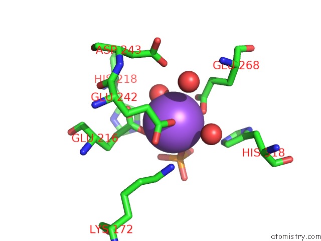
Mono view
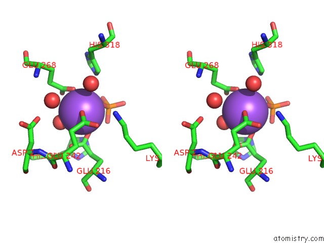
Stereo pair view
Mono view
Stereo pair view
A full contact list of Sodium with other atoms in the Na binding
site number 1 of Crystal Structure of An Enolase (Mandelate Racemase Subgroup, Target Efi-502086) From Agrobacterium Tumefaciens, with A Succinimide Residue, Na and Phosphate within 5.0Å range:
|
Reference:
M.W.Vetting,
J.T.Bouvier,
R.Toro,
R.Bhosle,
N.F.Al Obaidi,
L.L.Morisco,
S.R.Wasserman,
S.Sojitra,
H.J.Imker,
J.A.Gerlt,
S.C.Almo,
Enzyme Function Initiative (Efi).
Crystal Structure of An Enolase (Mandelate Racemase Subgroup, Target Efi-502086) From Agrobacterium Tumefaciens, with A Succinimide Residue, Na and Phosphate To Be Published.
Page generated: Mon Oct 7 15:01:16 2024
Last articles
F in 7NTHF in 7NTI
F in 7NPC
F in 7NRG
F in 7NR5
F in 7NQS
F in 7NOS
F in 7NP5
F in 7NDV
F in 7NP6