Sodium »
PDB 4def-4dxz »
4dn0 »
Sodium in PDB 4dn0: Peld 156-455 From Pseudomonas Aeruginosa PA14 in Complex with C-Di-Gmp
Protein crystallography data
The structure of Peld 156-455 From Pseudomonas Aeruginosa PA14 in Complex with C-Di-Gmp, PDB code: 4dn0
was solved by
J.C.Whitney,
K.M.Colvin,
L.S.Marmont,
H.Robinson,
M.R.Parsek,
P.L.Howell,
with X-Ray Crystallography technique. A brief refinement statistics is given in the table below:
| Resolution Low / High (Å) | 45.90 / 2.30 |
| Space group | P 21 21 21 |
| Cell size a, b, c (Å), α, β, γ (°) | 41.776, 82.043, 91.808, 90.00, 90.00, 90.00 |
| R / Rfree (%) | 20.1 / 25.2 |
Sodium Binding Sites:
The binding sites of Sodium atom in the Peld 156-455 From Pseudomonas Aeruginosa PA14 in Complex with C-Di-Gmp
(pdb code 4dn0). This binding sites where shown within
5.0 Angstroms radius around Sodium atom.
In total 2 binding sites of Sodium where determined in the Peld 156-455 From Pseudomonas Aeruginosa PA14 in Complex with C-Di-Gmp, PDB code: 4dn0:
Jump to Sodium binding site number: 1; 2;
In total 2 binding sites of Sodium where determined in the Peld 156-455 From Pseudomonas Aeruginosa PA14 in Complex with C-Di-Gmp, PDB code: 4dn0:
Jump to Sodium binding site number: 1; 2;
Sodium binding site 1 out of 2 in 4dn0
Go back to
Sodium binding site 1 out
of 2 in the Peld 156-455 From Pseudomonas Aeruginosa PA14 in Complex with C-Di-Gmp
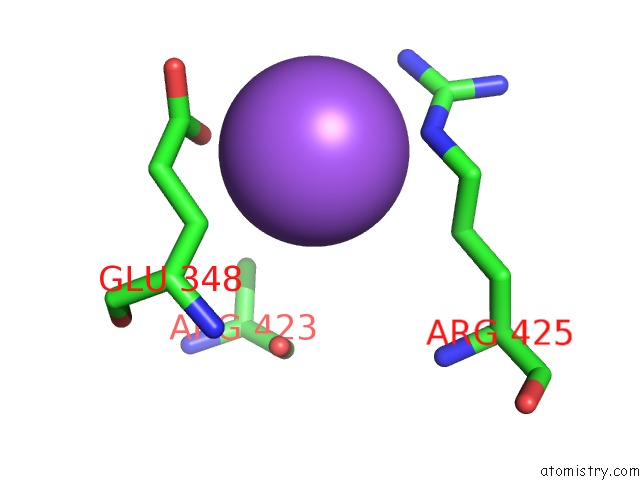
Mono view
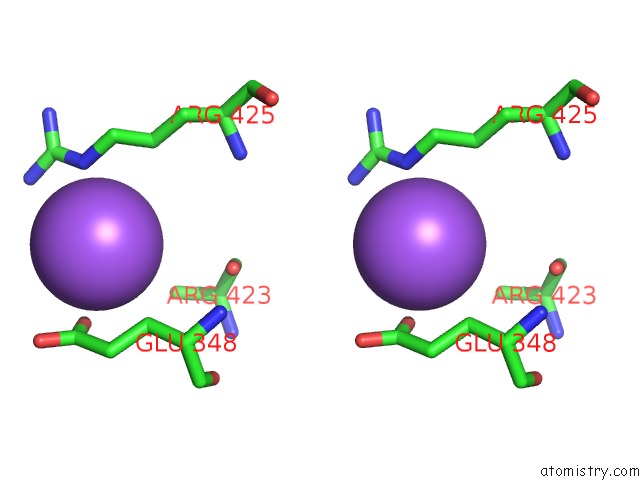
Stereo pair view
Mono view
Stereo pair view
A full contact list of Sodium with other atoms in the Na binding
site number 1 of Peld 156-455 From Pseudomonas Aeruginosa PA14 in Complex with C-Di-Gmp within 5.0Å range:
|
Sodium binding site 2 out of 2 in 4dn0
Go back to
Sodium binding site 2 out
of 2 in the Peld 156-455 From Pseudomonas Aeruginosa PA14 in Complex with C-Di-Gmp
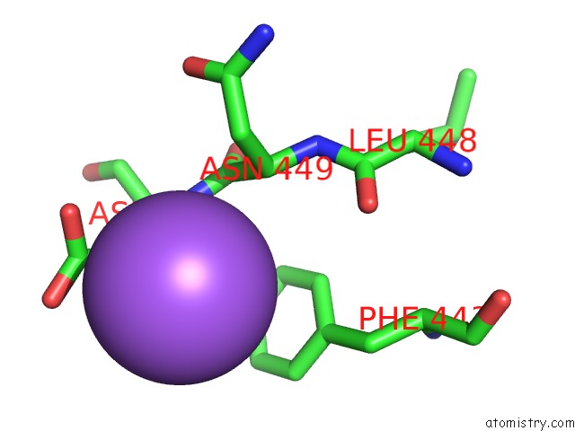
Mono view
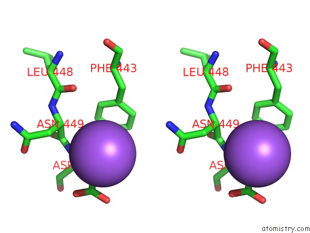
Stereo pair view
Mono view
Stereo pair view
A full contact list of Sodium with other atoms in the Na binding
site number 2 of Peld 156-455 From Pseudomonas Aeruginosa PA14 in Complex with C-Di-Gmp within 5.0Å range:
|
Reference:
J.C.Whitney,
K.M.Colvin,
L.S.Marmont,
H.Robinson,
M.R.Parsek,
P.L.Howell.
Structure of the Cytoplasmic Region of Peld, A Degenerate Diguanylate Cyclase Receptor That Regulates Exopolysaccharide Production in Pseudomonas Aeruginosa. J.Biol.Chem. V. 287 23582 2012.
ISSN: ISSN 0021-9258
PubMed: 22605337
DOI: 10.1074/JBC.M112.375378
Page generated: Mon Oct 7 14:57:40 2024
ISSN: ISSN 0021-9258
PubMed: 22605337
DOI: 10.1074/JBC.M112.375378
Last articles
Zn in 9JYWZn in 9IR4
Zn in 9IR3
Zn in 9GMX
Zn in 9GMW
Zn in 9JEJ
Zn in 9ERF
Zn in 9ERE
Zn in 9EGV
Zn in 9EGW