Sodium »
PDB 4blw-4c44 »
4brg »
Sodium in PDB 4brg: Legionella Pneumophila NTPDASE1 Crystal Form II (Closed) in Complex with Mg Gmppnp
Enzymatic activity of Legionella Pneumophila NTPDASE1 Crystal Form II (Closed) in Complex with Mg Gmppnp
All present enzymatic activity of Legionella Pneumophila NTPDASE1 Crystal Form II (Closed) in Complex with Mg Gmppnp:
3.6.1.5;
3.6.1.5;
Protein crystallography data
The structure of Legionella Pneumophila NTPDASE1 Crystal Form II (Closed) in Complex with Mg Gmppnp, PDB code: 4brg
was solved by
M.Zebisch,
P.Schaefer,
P.Lauble,
N.Straeter,
with X-Ray Crystallography technique. A brief refinement statistics is given in the table below:
| Resolution Low / High (Å) | 28.99 / 1.45 |
| Space group | P 1 21 1 |
| Cell size a, b, c (Å), α, β, γ (°) | 62.186, 86.166, 71.878, 90.00, 107.02, 90.00 |
| R / Rfree (%) | 12.371 / 17.444 |
Other elements in 4brg:
The structure of Legionella Pneumophila NTPDASE1 Crystal Form II (Closed) in Complex with Mg Gmppnp also contains other interesting chemical elements:
| Magnesium | (Mg) | 2 atoms |
| Chlorine | (Cl) | 2 atoms |
Sodium Binding Sites:
The binding sites of Sodium atom in the Legionella Pneumophila NTPDASE1 Crystal Form II (Closed) in Complex with Mg Gmppnp
(pdb code 4brg). This binding sites where shown within
5.0 Angstroms radius around Sodium atom.
In total only one binding site of Sodium was determined in the Legionella Pneumophila NTPDASE1 Crystal Form II (Closed) in Complex with Mg Gmppnp, PDB code: 4brg:
In total only one binding site of Sodium was determined in the Legionella Pneumophila NTPDASE1 Crystal Form II (Closed) in Complex with Mg Gmppnp, PDB code: 4brg:
Sodium binding site 1 out of 1 in 4brg
Go back to
Sodium binding site 1 out
of 1 in the Legionella Pneumophila NTPDASE1 Crystal Form II (Closed) in Complex with Mg Gmppnp
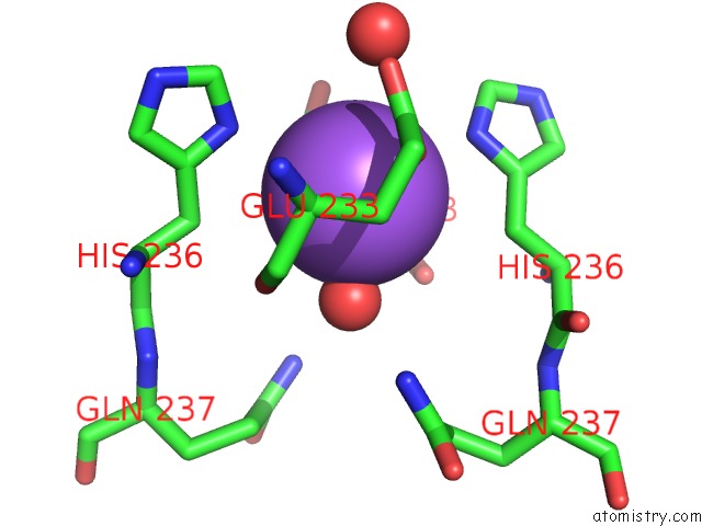
Mono view
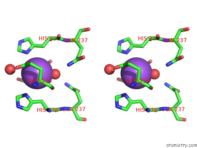
Stereo pair view
Mono view
Stereo pair view
A full contact list of Sodium with other atoms in the Na binding
site number 1 of Legionella Pneumophila NTPDASE1 Crystal Form II (Closed) in Complex with Mg Gmppnp within 5.0Å range:
|
Reference:
M.Zebisch,
M.Krauss,
P.Schaefer,
P.Lauble,
N.Straeter.
Crystallographic Snapshots Along the Reaction Pathway of Nucleoside Triphosphate Diphosphohydrolases Structure V. 21 1460 2013.
ISSN: ISSN 0969-2126
PubMed: 23830739
DOI: 10.1016/J.STR.2013.05.016
Page generated: Mon Oct 7 14:33:56 2024
ISSN: ISSN 0969-2126
PubMed: 23830739
DOI: 10.1016/J.STR.2013.05.016
Last articles
Ca in 5MLHCa in 5ML7
Ca in 5MKF
Ca in 5MKC
Ca in 5MIN
Ca in 5MKE
Ca in 5MJL
Ca in 5MIM
Ca in 5MIH
Ca in 5MJ7