Sodium »
PDB 4adn-4b16 »
4ayo »
Sodium in PDB 4ayo: Structure of the GH47 Processing Alpha-1,2-Mannosidase From Caulobacter Strain K31
Enzymatic activity of Structure of the GH47 Processing Alpha-1,2-Mannosidase From Caulobacter Strain K31
All present enzymatic activity of Structure of the GH47 Processing Alpha-1,2-Mannosidase From Caulobacter Strain K31:
3.2.1.113;
3.2.1.113;
Protein crystallography data
The structure of Structure of the GH47 Processing Alpha-1,2-Mannosidase From Caulobacter Strain K31, PDB code: 4ayo
was solved by
A.J.Thompson,
J.Dabin,
J.Iglesias-Fernandez,
A.Iglesias-Fernandez,
Z.Dinev,
S.J.Williams,
A.Siriwardena,
C.Moreland,
T.C.Hu,
D.K.Smith,
H.J.Gilbert,
C.Rovira,
G.J.Davies,
with X-Ray Crystallography technique. A brief refinement statistics is given in the table below:
| Resolution Low / High (Å) | 46.57 / 0.85 |
| Space group | H 3 |
| Cell size a, b, c (Å), α, β, γ (°) | 143.770, 143.770, 50.155, 90.00, 90.00, 120.00 |
| R / Rfree (%) | 9.464 / 10.478 |
Other elements in 4ayo:
The structure of Structure of the GH47 Processing Alpha-1,2-Mannosidase From Caulobacter Strain K31 also contains other interesting chemical elements:
| Calcium | (Ca) | 2 atoms |
Sodium Binding Sites:
The binding sites of Sodium atom in the Structure of the GH47 Processing Alpha-1,2-Mannosidase From Caulobacter Strain K31
(pdb code 4ayo). This binding sites where shown within
5.0 Angstroms radius around Sodium atom.
In total only one binding site of Sodium was determined in the Structure of the GH47 Processing Alpha-1,2-Mannosidase From Caulobacter Strain K31, PDB code: 4ayo:
In total only one binding site of Sodium was determined in the Structure of the GH47 Processing Alpha-1,2-Mannosidase From Caulobacter Strain K31, PDB code: 4ayo:
Sodium binding site 1 out of 1 in 4ayo
Go back to
Sodium binding site 1 out
of 1 in the Structure of the GH47 Processing Alpha-1,2-Mannosidase From Caulobacter Strain K31
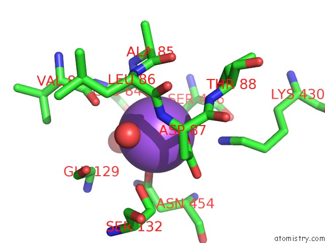
Mono view
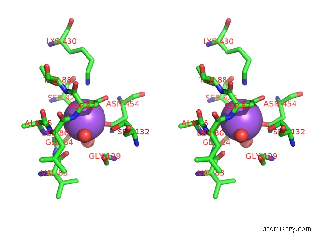
Stereo pair view
Mono view
Stereo pair view
A full contact list of Sodium with other atoms in the Na binding
site number 1 of Structure of the GH47 Processing Alpha-1,2-Mannosidase From Caulobacter Strain K31 within 5.0Å range:
|
Reference:
A.J.Thompson,
J.Dabin,
J.Iglesias-Fernandez,
A.Ardevol,
Z.Dinev,
S.J.Williams,
O.Bande,
A.Siriwardena,
C.Moreland,
T.C.Hu,
D.K.Smith,
H.J.Gilbert,
C.Rovira,
G.J.Davies.
The Reaction Coordinate of A Bacterial GH47 Alpha-Mannosidase: A Combined Quantum Mechanical and Structural Approach. Angew.Chem.Int.Ed.Engl. V. 51 10997 2012.
ISSN: ISSN 1433-7851
PubMed: 23012075
DOI: 10.1002/ANIE.201205338
Page generated: Mon Oct 7 14:25:10 2024
ISSN: ISSN 1433-7851
PubMed: 23012075
DOI: 10.1002/ANIE.201205338
Last articles
Cl in 5FJVCl in 5FKL
Cl in 5FJK
Cl in 5FJL
Cl in 5FJJ
Cl in 5FJG
Cl in 5FHO
Cl in 5FJH
Cl in 5FJ3
Cl in 5FIP