Sodium »
PDB 3wc3-3wxo »
3wky »
Sodium in PDB 3wky: Crystal Structure of Hemolymph Type Prophenoloxidase (Propob) From Crustacean
Protein crystallography data
The structure of Crystal Structure of Hemolymph Type Prophenoloxidase (Propob) From Crustacean, PDB code: 3wky
was solved by
T.Masuda,
B.Mikami,
with X-Ray Crystallography technique. A brief refinement statistics is given in the table below:
| Resolution Low / High (Å) | 34.81 / 1.80 |
| Space group | H 3 |
| Cell size a, b, c (Å), α, β, γ (°) | 156.706, 156.706, 283.830, 90.00, 90.00, 120.00 |
| R / Rfree (%) | 17.5 / 19.6 |
Other elements in 3wky:
The structure of Crystal Structure of Hemolymph Type Prophenoloxidase (Propob) From Crustacean also contains other interesting chemical elements:
| Magnesium | (Mg) | 1 atom |
| Copper | (Cu) | 4 atoms |
| Chlorine | (Cl) | 5 atoms |
Sodium Binding Sites:
The binding sites of Sodium atom in the Crystal Structure of Hemolymph Type Prophenoloxidase (Propob) From Crustacean
(pdb code 3wky). This binding sites where shown within
5.0 Angstroms radius around Sodium atom.
In total only one binding site of Sodium was determined in the Crystal Structure of Hemolymph Type Prophenoloxidase (Propob) From Crustacean, PDB code: 3wky:
In total only one binding site of Sodium was determined in the Crystal Structure of Hemolymph Type Prophenoloxidase (Propob) From Crustacean, PDB code: 3wky:
Sodium binding site 1 out of 1 in 3wky
Go back to
Sodium binding site 1 out
of 1 in the Crystal Structure of Hemolymph Type Prophenoloxidase (Propob) From Crustacean
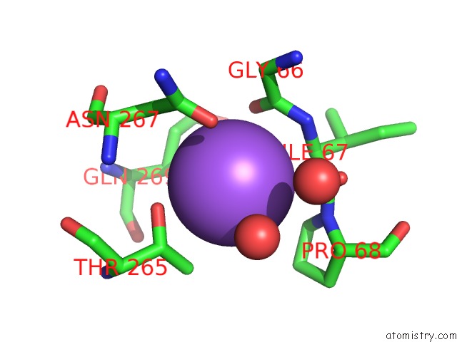
Mono view
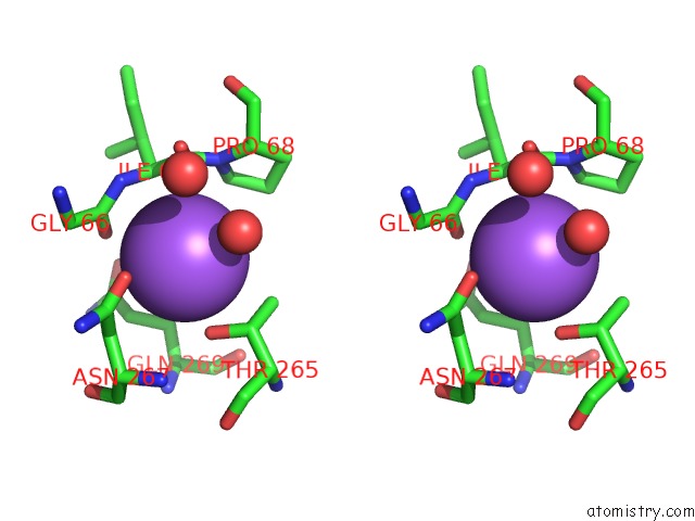
Stereo pair view
Mono view
Stereo pair view
A full contact list of Sodium with other atoms in the Na binding
site number 1 of Crystal Structure of Hemolymph Type Prophenoloxidase (Propob) From Crustacean within 5.0Å range:
|
Reference:
T.Masuda,
K.Momoji,
T.Hirata,
B.Mikami.
Crystal Structure of A Crustacean Prophenoloxidase Provides A Clue to Understanding the Functionality of the Type 3 Copper Proteins. Febs J. 2014.
ISSN: ISSN 1742-464X
PubMed: 24720693
DOI: 10.1111/FEBS.12812
Page generated: Mon Oct 7 13:58:07 2024
ISSN: ISSN 1742-464X
PubMed: 24720693
DOI: 10.1111/FEBS.12812
Last articles
Cl in 5GVMCl in 5GTY
Cl in 5GVL
Cl in 5GVK
Cl in 5GUS
Cl in 5GUW
Cl in 5GUK
Cl in 5GTZ
Cl in 5GU6
Cl in 5GTI