Sodium »
PDB 3wc3-3wxo »
3we7 »
Sodium in PDB 3we7: Crystal Structure of Diacetylchitobiose Deacetylase From Pyrococcus Horikoshii
Protein crystallography data
The structure of Crystal Structure of Diacetylchitobiose Deacetylase From Pyrococcus Horikoshii, PDB code: 3we7
was solved by
S.Mine,
T.Nakamura,
Y.Fukuda,
T.Inoue,
K.Uegaki,
T.Sato,
with X-Ray Crystallography technique. A brief refinement statistics is given in the table below:
| Resolution Low / High (Å) | 29.62 / 1.55 |
| Space group | P 32 2 1 |
| Cell size a, b, c (Å), α, β, γ (°) | 77.372, 77.372, 230.223, 90.00, 90.00, 120.00 |
| R / Rfree (%) | 17.3 / 20.7 |
Other elements in 3we7:
The structure of Crystal Structure of Diacetylchitobiose Deacetylase From Pyrococcus Horikoshii also contains other interesting chemical elements:
| Zinc | (Zn) | 3 atoms |
Sodium Binding Sites:
The binding sites of Sodium atom in the Crystal Structure of Diacetylchitobiose Deacetylase From Pyrococcus Horikoshii
(pdb code 3we7). This binding sites where shown within
5.0 Angstroms radius around Sodium atom.
In total 2 binding sites of Sodium where determined in the Crystal Structure of Diacetylchitobiose Deacetylase From Pyrococcus Horikoshii, PDB code: 3we7:
Jump to Sodium binding site number: 1; 2;
In total 2 binding sites of Sodium where determined in the Crystal Structure of Diacetylchitobiose Deacetylase From Pyrococcus Horikoshii, PDB code: 3we7:
Jump to Sodium binding site number: 1; 2;
Sodium binding site 1 out of 2 in 3we7
Go back to
Sodium binding site 1 out
of 2 in the Crystal Structure of Diacetylchitobiose Deacetylase From Pyrococcus Horikoshii
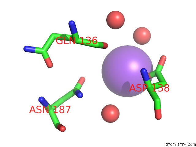
Mono view
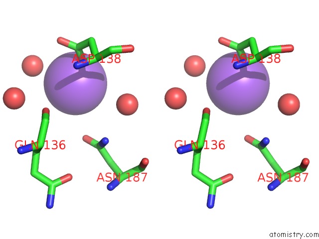
Stereo pair view
Mono view
Stereo pair view
A full contact list of Sodium with other atoms in the Na binding
site number 1 of Crystal Structure of Diacetylchitobiose Deacetylase From Pyrococcus Horikoshii within 5.0Å range:
|
Sodium binding site 2 out of 2 in 3we7
Go back to
Sodium binding site 2 out
of 2 in the Crystal Structure of Diacetylchitobiose Deacetylase From Pyrococcus Horikoshii
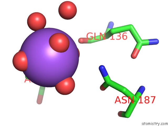
Mono view
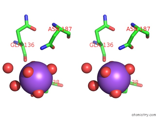
Stereo pair view
Mono view
Stereo pair view
A full contact list of Sodium with other atoms in the Na binding
site number 2 of Crystal Structure of Diacetylchitobiose Deacetylase From Pyrococcus Horikoshii within 5.0Å range:
|
Reference:
S.Mine,
M.Niiyama,
W.Hashimoto,
T.Ikegami,
D.Koma,
T.Ohmoto,
Y.Fukuda,
T.Inoue,
Y.Abe,
T.Ueda,
J.Morita,
K.Uegaki,
T.Nakamura.
Expression From Engineered Escherichia Coli Chromosome and Crystallographic Study of Archaeal N,N'-Diacetylchitobiose Deacetylase Febs J. V. 281 2584 2014.
ISSN: ISSN 1742-464X
PubMed: 24702737
DOI: 10.1111/FEBS.12805
Page generated: Mon Oct 7 13:56:40 2024
ISSN: ISSN 1742-464X
PubMed: 24702737
DOI: 10.1111/FEBS.12805
Last articles
Cl in 5TERCl in 5TEX
Cl in 5TEK
Cl in 5TE8
Cl in 5TEL
Cl in 5TEI
Cl in 5TD5
Cl in 5TD9
Cl in 5TDS
Cl in 5T9P