Sodium »
PDB 3wc3-3wxo »
3wdr »
Sodium in PDB 3wdr: Crystal Structure of Beta-Mannanase From A Symbiotic Protist of the Termite Reticulitermes Speratus Complexed with Gluco-Manno- Oligosaccharide
Enzymatic activity of Crystal Structure of Beta-Mannanase From A Symbiotic Protist of the Termite Reticulitermes Speratus Complexed with Gluco-Manno- Oligosaccharide
All present enzymatic activity of Crystal Structure of Beta-Mannanase From A Symbiotic Protist of the Termite Reticulitermes Speratus Complexed with Gluco-Manno- Oligosaccharide:
3.2.1.78;
3.2.1.78;
Protein crystallography data
The structure of Crystal Structure of Beta-Mannanase From A Symbiotic Protist of the Termite Reticulitermes Speratus Complexed with Gluco-Manno- Oligosaccharide, PDB code: 3wdr
was solved by
H.Tsukagoshi,
T.Ishida,
K.K.Touhara,
K.Igarashi,
M.Samejima,
S.Fushinobu,
K.Kitamoto,
M.Arioka,
with X-Ray Crystallography technique. A brief refinement statistics is given in the table below:
| Resolution Low / High (Å) | 36.51 / 1.40 |
| Space group | P 1 21 1 |
| Cell size a, b, c (Å), α, β, γ (°) | 45.522, 61.644, 58.068, 90.00, 92.27, 90.00 |
| R / Rfree (%) | 13.7 / 16 |
Other elements in 3wdr:
The structure of Crystal Structure of Beta-Mannanase From A Symbiotic Protist of the Termite Reticulitermes Speratus Complexed with Gluco-Manno- Oligosaccharide also contains other interesting chemical elements:
| Magnesium | (Mg) | 2 atoms |
Sodium Binding Sites:
The binding sites of Sodium atom in the Crystal Structure of Beta-Mannanase From A Symbiotic Protist of the Termite Reticulitermes Speratus Complexed with Gluco-Manno- Oligosaccharide
(pdb code 3wdr). This binding sites where shown within
5.0 Angstroms radius around Sodium atom.
In total 2 binding sites of Sodium where determined in the Crystal Structure of Beta-Mannanase From A Symbiotic Protist of the Termite Reticulitermes Speratus Complexed with Gluco-Manno- Oligosaccharide, PDB code: 3wdr:
Jump to Sodium binding site number: 1; 2;
In total 2 binding sites of Sodium where determined in the Crystal Structure of Beta-Mannanase From A Symbiotic Protist of the Termite Reticulitermes Speratus Complexed with Gluco-Manno- Oligosaccharide, PDB code: 3wdr:
Jump to Sodium binding site number: 1; 2;
Sodium binding site 1 out of 2 in 3wdr
Go back to
Sodium binding site 1 out
of 2 in the Crystal Structure of Beta-Mannanase From A Symbiotic Protist of the Termite Reticulitermes Speratus Complexed with Gluco-Manno- Oligosaccharide
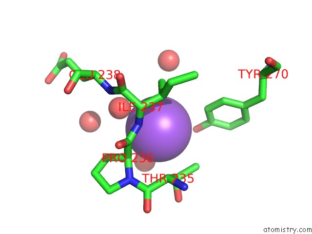
Mono view
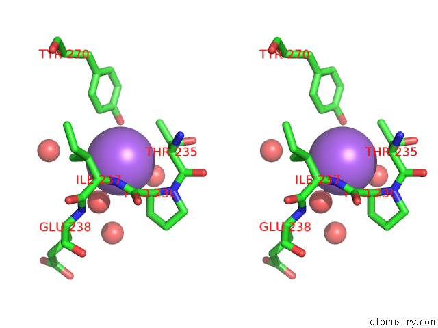
Stereo pair view
Mono view
Stereo pair view
A full contact list of Sodium with other atoms in the Na binding
site number 1 of Crystal Structure of Beta-Mannanase From A Symbiotic Protist of the Termite Reticulitermes Speratus Complexed with Gluco-Manno- Oligosaccharide within 5.0Å range:
|
Sodium binding site 2 out of 2 in 3wdr
Go back to
Sodium binding site 2 out
of 2 in the Crystal Structure of Beta-Mannanase From A Symbiotic Protist of the Termite Reticulitermes Speratus Complexed with Gluco-Manno- Oligosaccharide
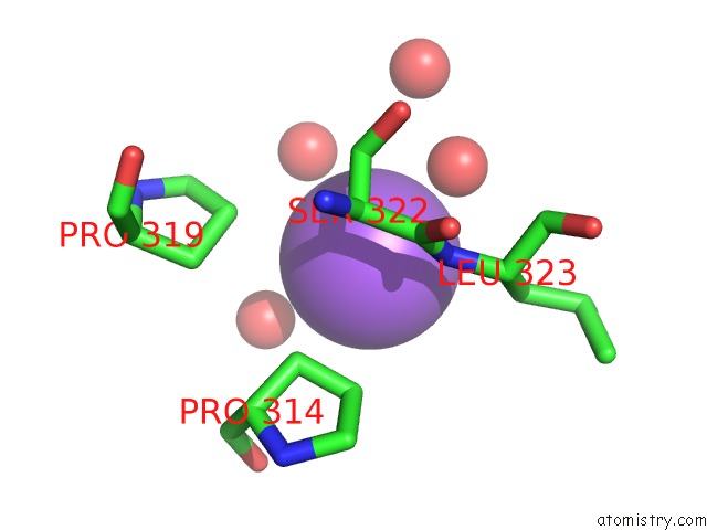
Mono view
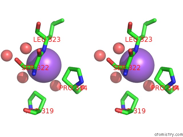
Stereo pair view
Mono view
Stereo pair view
A full contact list of Sodium with other atoms in the Na binding
site number 2 of Crystal Structure of Beta-Mannanase From A Symbiotic Protist of the Termite Reticulitermes Speratus Complexed with Gluco-Manno- Oligosaccharide within 5.0Å range:
|
Reference:
H.Tsukagoshi,
A.Nakamura,
T.Ishida,
K.K.Touhara,
M.Otagiri,
S.Moriya,
M.Samejima,
K.Igarashi,
S.Fushinobu,
K.Kitamoto,
M.Arioka.
Structural and Biochemical Analyses of Glycoside Hydrolase Family 26 Beta-Mannanase From A Symbiotic Protist of the Termite Reticulitermes Speratus J.Biol.Chem. V. 289 10843 2014.
ISSN: ISSN 0021-9258
PubMed: 24570006
DOI: 10.1074/JBC.M114.555383
Page generated: Mon Oct 7 13:56:40 2024
ISSN: ISSN 0021-9258
PubMed: 24570006
DOI: 10.1074/JBC.M114.555383
Last articles
Zn in 9J0NZn in 9J0O
Zn in 9J0P
Zn in 9FJX
Zn in 9EKB
Zn in 9C0F
Zn in 9CAH
Zn in 9CH0
Zn in 9CH3
Zn in 9CH1