Sodium »
PDB 3wc3-3wxo »
3wc3 »
Sodium in PDB 3wc3: Crystal Structure of Endo-1,4-Beta-Glucanase From Eisenia Fetida
Enzymatic activity of Crystal Structure of Endo-1,4-Beta-Glucanase From Eisenia Fetida
All present enzymatic activity of Crystal Structure of Endo-1,4-Beta-Glucanase From Eisenia Fetida:
3.2.1.4;
3.2.1.4;
Protein crystallography data
The structure of Crystal Structure of Endo-1,4-Beta-Glucanase From Eisenia Fetida, PDB code: 3wc3
was solved by
T.Arimori,
T.Tamada,
with X-Ray Crystallography technique. A brief refinement statistics is given in the table below:
| Resolution Low / High (Å) | 28.20 / 1.50 |
| Space group | P 32 2 1 |
| Cell size a, b, c (Å), α, β, γ (°) | 136.449, 136.449, 54.974, 90.00, 90.00, 120.00 |
| R / Rfree (%) | 14.7 / 16.8 |
Other elements in 3wc3:
The structure of Crystal Structure of Endo-1,4-Beta-Glucanase From Eisenia Fetida also contains other interesting chemical elements:
| Calcium | (Ca) | 1 atom |
Sodium Binding Sites:
The binding sites of Sodium atom in the Crystal Structure of Endo-1,4-Beta-Glucanase From Eisenia Fetida
(pdb code 3wc3). This binding sites where shown within
5.0 Angstroms radius around Sodium atom.
In total only one binding site of Sodium was determined in the Crystal Structure of Endo-1,4-Beta-Glucanase From Eisenia Fetida, PDB code: 3wc3:
In total only one binding site of Sodium was determined in the Crystal Structure of Endo-1,4-Beta-Glucanase From Eisenia Fetida, PDB code: 3wc3:
Sodium binding site 1 out of 1 in 3wc3
Go back to
Sodium binding site 1 out
of 1 in the Crystal Structure of Endo-1,4-Beta-Glucanase From Eisenia Fetida
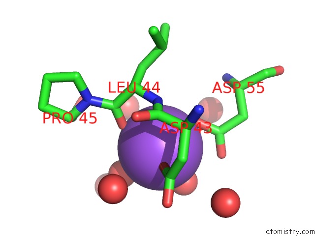
Mono view
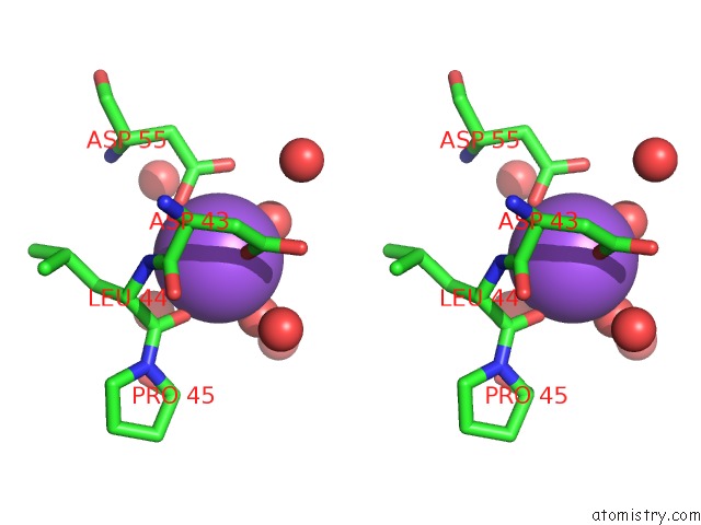
Stereo pair view
Mono view
Stereo pair view
A full contact list of Sodium with other atoms in the Na binding
site number 1 of Crystal Structure of Endo-1,4-Beta-Glucanase From Eisenia Fetida within 5.0Å range:
|
Reference:
T.Arimori,
A.Ito,
M.Nakazawa,
M.Ueda,
T.Tamada.
Crystal Structure of Endo-1,4-Beta-Glucanase From Eisenia Fetida J.Synchrotron Radiat. V. 20 884 2013.
ISSN: ISSN 0909-0495
PubMed: 24121333
DOI: 10.1107/S0909049513021110
Page generated: Mon Oct 7 13:56:40 2024
ISSN: ISSN 0909-0495
PubMed: 24121333
DOI: 10.1107/S0909049513021110
Last articles
Zn in 9MJ5Zn in 9HNW
Zn in 9G0L
Zn in 9FNE
Zn in 9DZN
Zn in 9E0I
Zn in 9D32
Zn in 9DAK
Zn in 8ZXC
Zn in 8ZUF