Sodium »
PDB 3sib-3t09 »
3sya »
Sodium in PDB 3sya: Crystal Structure of the G Protein-Gated Inward Rectifier K+ Channel GIRK2 (KIR3.2) in Complex with Sodium and PIP2
Protein crystallography data
The structure of Crystal Structure of the G Protein-Gated Inward Rectifier K+ Channel GIRK2 (KIR3.2) in Complex with Sodium and PIP2, PDB code: 3sya
was solved by
M.R.Whorton,
R.Mackinnon,
with X-Ray Crystallography technique. A brief refinement statistics is given in the table below:
| Resolution Low / High (Å) | 41.73 / 2.98 |
| Space group | P 4 21 2 |
| Cell size a, b, c (Å), α, β, γ (°) | 85.851, 85.851, 177.862, 90.00, 90.00, 90.00 |
| R / Rfree (%) | 24 / 26.9 |
Other elements in 3sya:
The structure of Crystal Structure of the G Protein-Gated Inward Rectifier K+ Channel GIRK2 (KIR3.2) in Complex with Sodium and PIP2 also contains other interesting chemical elements:
| Potassium | (K) | 5 atoms |
Sodium Binding Sites:
The binding sites of Sodium atom in the Crystal Structure of the G Protein-Gated Inward Rectifier K+ Channel GIRK2 (KIR3.2) in Complex with Sodium and PIP2
(pdb code 3sya). This binding sites where shown within
5.0 Angstroms radius around Sodium atom.
In total only one binding site of Sodium was determined in the Crystal Structure of the G Protein-Gated Inward Rectifier K+ Channel GIRK2 (KIR3.2) in Complex with Sodium and PIP2, PDB code: 3sya:
In total only one binding site of Sodium was determined in the Crystal Structure of the G Protein-Gated Inward Rectifier K+ Channel GIRK2 (KIR3.2) in Complex with Sodium and PIP2, PDB code: 3sya:
Sodium binding site 1 out of 1 in 3sya
Go back to
Sodium binding site 1 out
of 1 in the Crystal Structure of the G Protein-Gated Inward Rectifier K+ Channel GIRK2 (KIR3.2) in Complex with Sodium and PIP2
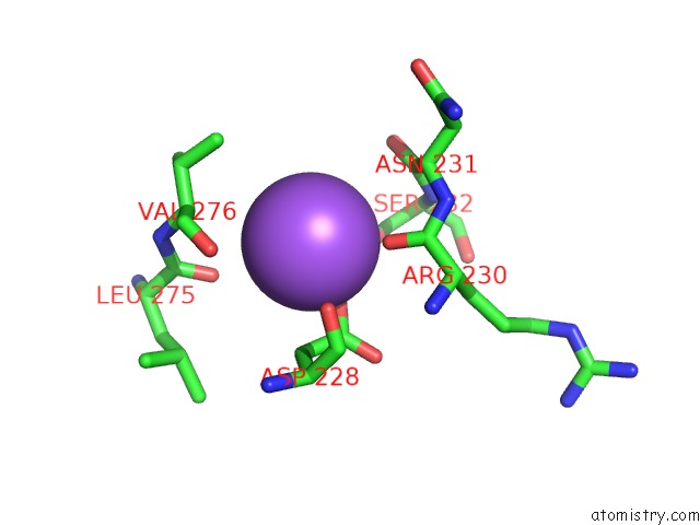
Mono view
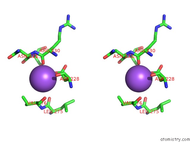
Stereo pair view
Mono view
Stereo pair view
A full contact list of Sodium with other atoms in the Na binding
site number 1 of Crystal Structure of the G Protein-Gated Inward Rectifier K+ Channel GIRK2 (KIR3.2) in Complex with Sodium and PIP2 within 5.0Å range:
|
Reference:
M.R.Whorton,
R.Mackinnon.
Crystal Structure of the Mammalian GIRK2 K(+) Channel and Gating Regulation By G Proteins, Pip(2), and Sodium. Cell(Cambridge,Mass.) V. 147 199 2011.
ISSN: ISSN 0092-8674
PubMed: 21962516
DOI: 10.1016/J.CELL.2011.07.046
Page generated: Mon Oct 7 13:01:51 2024
ISSN: ISSN 0092-8674
PubMed: 21962516
DOI: 10.1016/J.CELL.2011.07.046
Last articles
Cl in 5TM9Cl in 5TML
Cl in 5TNO
Cl in 5TM7
Cl in 5TM4
Cl in 5TM3
Cl in 5TL4
Cl in 5TM2
Cl in 5TLL
Cl in 5TLS