Sodium »
PDB 3sib-3t09 »
3svi »
Sodium in PDB 3svi: Structure of the Pto-Binding Domain of Hoppmal Generated By Limited Thermolysin Digestion
Protein crystallography data
The structure of Structure of the Pto-Binding Domain of Hoppmal Generated By Limited Thermolysin Digestion, PDB code: 3svi
was solved by
A.U.Singer,
A.Stein,
X.Xu,
H.Cui,
A.Joachimiak,
A.M.Edwards,
A.Savchenko,
Midwest Center For Structural Genomics (Mcsg),
with X-Ray Crystallography technique. A brief refinement statistics is given in the table below:
| Resolution Low / High (Å) | 25.62 / 1.80 |
| Space group | P 41 21 2 |
| Cell size a, b, c (Å), α, β, γ (°) | 57.277, 57.277, 55.658, 90.00, 90.00, 90.00 |
| R / Rfree (%) | 18.6 / 21.8 |
Other elements in 3svi:
The structure of Structure of the Pto-Binding Domain of Hoppmal Generated By Limited Thermolysin Digestion also contains other interesting chemical elements:
| Chlorine | (Cl) | 4 atoms |
Sodium Binding Sites:
The binding sites of Sodium atom in the Structure of the Pto-Binding Domain of Hoppmal Generated By Limited Thermolysin Digestion
(pdb code 3svi). This binding sites where shown within
5.0 Angstroms radius around Sodium atom.
In total only one binding site of Sodium was determined in the Structure of the Pto-Binding Domain of Hoppmal Generated By Limited Thermolysin Digestion, PDB code: 3svi:
In total only one binding site of Sodium was determined in the Structure of the Pto-Binding Domain of Hoppmal Generated By Limited Thermolysin Digestion, PDB code: 3svi:
Sodium binding site 1 out of 1 in 3svi
Go back to
Sodium binding site 1 out
of 1 in the Structure of the Pto-Binding Domain of Hoppmal Generated By Limited Thermolysin Digestion
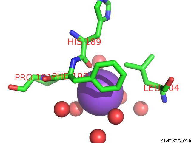
Mono view
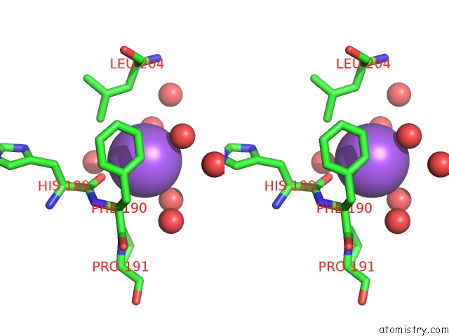
Stereo pair view
Mono view
Stereo pair view
A full contact list of Sodium with other atoms in the Na binding
site number 1 of Structure of the Pto-Binding Domain of Hoppmal Generated By Limited Thermolysin Digestion within 5.0Å range:
|
Reference:
A.U.Singer,
B.Wu,
A.Yee,
S.Houliston,
X.Xu,
H.Cui,
T.Skarina,
M.Garcia,
A.Semesi,
C.H.Arrowsmith,
A.Savchenko.
Structural Analysis of Hoppmal Reveals the Presence of A Second Adaptor Domain Common to the Hopab Family of Pseudomonas Syringae Type III Effectors. Biochemistry V. 51 1 2012.
ISSN: ISSN 0006-2960
PubMed: 22191472
DOI: 10.1021/BI2013883
Page generated: Mon Oct 7 13:00:58 2024
ISSN: ISSN 0006-2960
PubMed: 22191472
DOI: 10.1021/BI2013883
Last articles
Zn in 9J0NZn in 9J0O
Zn in 9J0P
Zn in 9FJX
Zn in 9EKB
Zn in 9C0F
Zn in 9CAH
Zn in 9CH0
Zn in 9CH3
Zn in 9CH1