Sodium »
PDB 3sib-3t09 »
3slz »
Sodium in PDB 3slz: The Crystal Structure of Xmrv Protease Complexed with Tl-3
Protein crystallography data
The structure of The Crystal Structure of Xmrv Protease Complexed with Tl-3, PDB code: 3slz
was solved by
M.Li,
A.Gustchina,
A.Wlodawer,
with X-Ray Crystallography technique. A brief refinement statistics is given in the table below:
| Resolution Low / High (Å) | 47.78 / 1.40 |
| Space group | P 21 21 21 |
| Cell size a, b, c (Å), α, β, γ (°) | 46.483, 65.546, 69.732, 90.00, 90.00, 90.00 |
| R / Rfree (%) | 17.6 / 20.1 |
Sodium Binding Sites:
The binding sites of Sodium atom in the The Crystal Structure of Xmrv Protease Complexed with Tl-3
(pdb code 3slz). This binding sites where shown within
5.0 Angstroms radius around Sodium atom.
In total only one binding site of Sodium was determined in the The Crystal Structure of Xmrv Protease Complexed with Tl-3, PDB code: 3slz:
In total only one binding site of Sodium was determined in the The Crystal Structure of Xmrv Protease Complexed with Tl-3, PDB code: 3slz:
Sodium binding site 1 out of 1 in 3slz
Go back to
Sodium binding site 1 out
of 1 in the The Crystal Structure of Xmrv Protease Complexed with Tl-3

Mono view
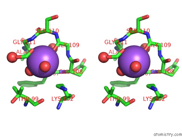
Stereo pair view
Mono view
Stereo pair view
A full contact list of Sodium with other atoms in the Na binding
site number 1 of The Crystal Structure of Xmrv Protease Complexed with Tl-3 within 5.0Å range:
|
Reference:
M.Li,
A.Gustchina,
K.Matuz,
J.Tozser,
S.Namwong,
N.E.Goldfarb,
B.M.Dunn,
A.Wlodawer.
Structural and Biochemical Characterization of the Inhibitor Complexes of Xenotropic Murine Leukemia Virus-Related Virus Protease. Febs J. V. 278 4413 2011.
ISSN: ISSN 1742-464X
PubMed: 21951660
DOI: 10.1111/J.1742-4658.2011.08364.X
Page generated: Sun Aug 17 17:23:24 2025
ISSN: ISSN 1742-464X
PubMed: 21951660
DOI: 10.1111/J.1742-4658.2011.08364.X
Last articles
Na in 5NJRNa in 5NJQ
Na in 5NJP
Na in 5NJC
Na in 5NI3
Na in 5NJA
Na in 5NJ9
Na in 5NJ1
Na in 5NII
Na in 5NIB