Sodium »
PDB 3sib-3t09 »
3sla »
Sodium in PDB 3sla: X-Ray Structure of First Four Repeats of Human Beta-Catenin
Protein crystallography data
The structure of X-Ray Structure of First Four Repeats of Human Beta-Catenin, PDB code: 3sla
was solved by
D.Gupta,
M.Bienz,
with X-Ray Crystallography technique. A brief refinement statistics is given in the table below:
| Resolution Low / High (Å) | 40.71 / 2.50 |
| Space group | P 41 21 2 |
| Cell size a, b, c (Å), α, β, γ (°) | 90.790, 90.790, 364.340, 90.00, 90.00, 90.00 |
| R / Rfree (%) | 21.8 / 27.7 |
Sodium Binding Sites:
The binding sites of Sodium atom in the X-Ray Structure of First Four Repeats of Human Beta-Catenin
(pdb code 3sla). This binding sites where shown within
5.0 Angstroms radius around Sodium atom.
In total 2 binding sites of Sodium where determined in the X-Ray Structure of First Four Repeats of Human Beta-Catenin, PDB code: 3sla:
Jump to Sodium binding site number: 1; 2;
In total 2 binding sites of Sodium where determined in the X-Ray Structure of First Four Repeats of Human Beta-Catenin, PDB code: 3sla:
Jump to Sodium binding site number: 1; 2;
Sodium binding site 1 out of 2 in 3sla
Go back to
Sodium binding site 1 out
of 2 in the X-Ray Structure of First Four Repeats of Human Beta-Catenin
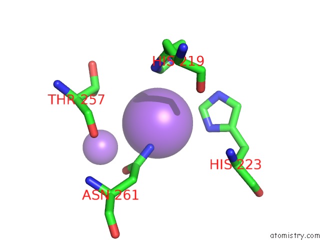
Mono view
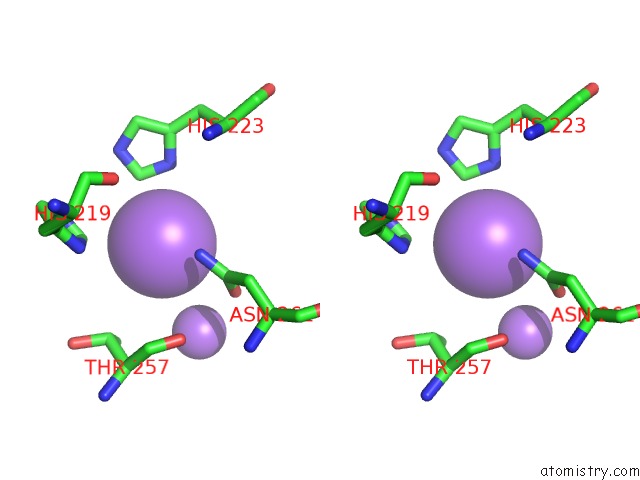
Stereo pair view
Mono view
Stereo pair view
A full contact list of Sodium with other atoms in the Na binding
site number 1 of X-Ray Structure of First Four Repeats of Human Beta-Catenin within 5.0Å range:
|
Sodium binding site 2 out of 2 in 3sla
Go back to
Sodium binding site 2 out
of 2 in the X-Ray Structure of First Four Repeats of Human Beta-Catenin
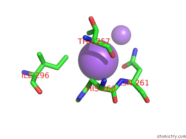
Mono view
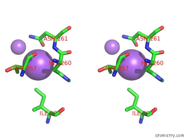
Stereo pair view
Mono view
Stereo pair view
A full contact list of Sodium with other atoms in the Na binding
site number 2 of X-Ray Structure of First Four Repeats of Human Beta-Catenin within 5.0Å range:
|
Reference:
M.De La Roche,
T.J.Rutherford,
D.Gupta,
D.B.Veprintsev,
B.Saxty,
S.M.Freund,
M.Bienz.
An Intrinsically Labile Alpha-Helix Abutting the BCL9-Binding Site of Beta-Catenin Is Required For Its Inhibition By Carnosic Acid. Nat Commun V. 3 680 2012.
ISSN: ESSN 2041-1723
PubMed: 22353711
DOI: 10.1038/NCOMMS1680
Page generated: Sun Aug 17 17:23:23 2025
ISSN: ESSN 2041-1723
PubMed: 22353711
DOI: 10.1038/NCOMMS1680
Last articles
Na in 7A2KNa in 7A36
Na in 7A2M
Na in 7A33
Na in 7A20
Na in 7A2L
Na in 7A2J
Na in 7A1I
Na in 7A1R
Na in 7A0S