Sodium »
PDB 3iwf-3k93 »
3jw2 »
Sodium in PDB 3jw2: Hiv-1 Protease Mutant G86S with Darunavir
Enzymatic activity of Hiv-1 Protease Mutant G86S with Darunavir
All present enzymatic activity of Hiv-1 Protease Mutant G86S with Darunavir:
3.4.23.16;
3.4.23.16;
Protein crystallography data
The structure of Hiv-1 Protease Mutant G86S with Darunavir, PDB code: 3jw2
was solved by
Y.Tie,
I.T.Weber,
with X-Ray Crystallography technique. A brief refinement statistics is given in the table below:
| Resolution Low / High (Å) | 10.00 / 1.80 |
| Space group | P 21 21 2 |
| Cell size a, b, c (Å), α, β, γ (°) | 58.355, 86.145, 46.369, 90.00, 90.00, 90.00 |
| R / Rfree (%) | 19.6 / 28.2 |
Other elements in 3jw2:
The structure of Hiv-1 Protease Mutant G86S with Darunavir also contains other interesting chemical elements:
| Chlorine | (Cl) | 1 atom |
Sodium Binding Sites:
The binding sites of Sodium atom in the Hiv-1 Protease Mutant G86S with Darunavir
(pdb code 3jw2). This binding sites where shown within
5.0 Angstroms radius around Sodium atom.
In total only one binding site of Sodium was determined in the Hiv-1 Protease Mutant G86S with Darunavir, PDB code: 3jw2:
In total only one binding site of Sodium was determined in the Hiv-1 Protease Mutant G86S with Darunavir, PDB code: 3jw2:
Sodium binding site 1 out of 1 in 3jw2
Go back to
Sodium binding site 1 out
of 1 in the Hiv-1 Protease Mutant G86S with Darunavir
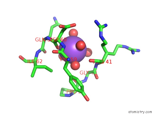
Mono view
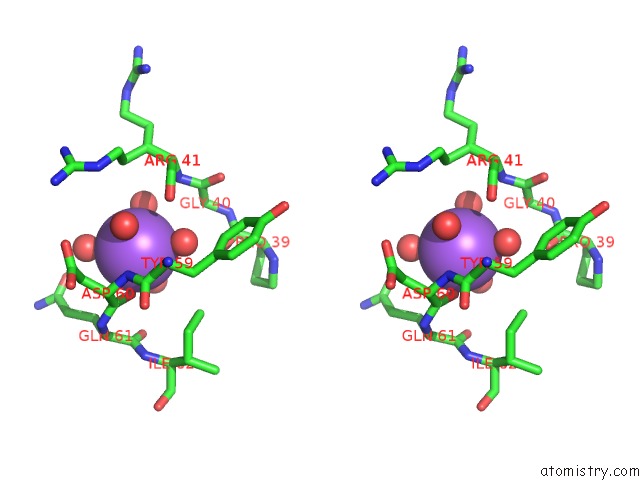
Stereo pair view
Mono view
Stereo pair view
A full contact list of Sodium with other atoms in the Na binding
site number 1 of Hiv-1 Protease Mutant G86S with Darunavir within 5.0Å range:
|
Reference:
R.Ishima,
Q.Gong,
Y.Tie,
I.T.Weber,
J.M.Louis.
Highly Conserved Glycine 86 and Arginine 87 Residues Contribute Differently to the Structure and Activity of the Mature Hiv-1 Protease Proteins V. 78 1015 2009.
ISSN: ISSN 0887-3585
PubMed: 19899162
DOI: 10.1002/PROT.22625
Page generated: Mon Oct 7 11:06:29 2024
ISSN: ISSN 0887-3585
PubMed: 19899162
DOI: 10.1002/PROT.22625
Last articles
Zn in 9MJ5Zn in 9HNW
Zn in 9G0L
Zn in 9FNE
Zn in 9DZN
Zn in 9E0I
Zn in 9D32
Zn in 9DAK
Zn in 8ZXC
Zn in 8ZUF