Sodium »
PDB 3iwf-3k93 »
3js5 »
Sodium in PDB 3js5: Crystal Structure of Protein Tyrosine Phosphatase From Entamoeba Histolytica with Hepes in the Active Site. High Resolution, Alternative Crystal Form with 1 Molecule in Asymmetric Unit
Protein crystallography data
The structure of Crystal Structure of Protein Tyrosine Phosphatase From Entamoeba Histolytica with Hepes in the Active Site. High Resolution, Alternative Crystal Form with 1 Molecule in Asymmetric Unit, PDB code: 3js5
was solved by
Seattle Structural Genomics Center For Infectious Disease (Ssgcid),
with X-Ray Crystallography technique. A brief refinement statistics is given in the table below:
| Resolution Low / High (Å) | 46.24 / 1.94 |
| Space group | P 21 21 21 |
| Cell size a, b, c (Å), α, β, γ (°) | 38.433, 60.195, 72.229, 90.00, 90.00, 90.00 |
| R / Rfree (%) | 16.7 / 21.2 |
Sodium Binding Sites:
The binding sites of Sodium atom in the Crystal Structure of Protein Tyrosine Phosphatase From Entamoeba Histolytica with Hepes in the Active Site. High Resolution, Alternative Crystal Form with 1 Molecule in Asymmetric Unit
(pdb code 3js5). This binding sites where shown within
5.0 Angstroms radius around Sodium atom.
In total only one binding site of Sodium was determined in the Crystal Structure of Protein Tyrosine Phosphatase From Entamoeba Histolytica with Hepes in the Active Site. High Resolution, Alternative Crystal Form with 1 Molecule in Asymmetric Unit, PDB code: 3js5:
In total only one binding site of Sodium was determined in the Crystal Structure of Protein Tyrosine Phosphatase From Entamoeba Histolytica with Hepes in the Active Site. High Resolution, Alternative Crystal Form with 1 Molecule in Asymmetric Unit, PDB code: 3js5:
Sodium binding site 1 out of 1 in 3js5
Go back to
Sodium binding site 1 out
of 1 in the Crystal Structure of Protein Tyrosine Phosphatase From Entamoeba Histolytica with Hepes in the Active Site. High Resolution, Alternative Crystal Form with 1 Molecule in Asymmetric Unit
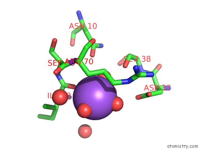
Mono view
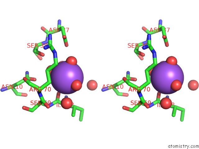
Stereo pair view
Mono view
Stereo pair view
A full contact list of Sodium with other atoms in the Na binding
site number 1 of Crystal Structure of Protein Tyrosine Phosphatase From Entamoeba Histolytica with Hepes in the Active Site. High Resolution, Alternative Crystal Form with 1 Molecule in Asymmetric Unit within 5.0Å range:
|
Reference:
A.S.Linford,
N.M.Jiang,
T.E.Edwards,
N.E.Sherman,
W.C.Van Voorhis,
L.J.Stewart,
P.J.Myler,
B.L.Staker,
W.A.Petri.
Crystal Structure and Putative Substrate Identification For the Entamoeba Histolytica Low Molecular Weight Tyrosine Phosphatase. Mol.Biochem.Parasitol. V. 193 33 2014.
ISSN: ISSN 0166-6851
PubMed: 24548880
DOI: 10.1016/J.MOLBIOPARA.2014.01.003
Page generated: Mon Oct 7 11:06:23 2024
ISSN: ISSN 0166-6851
PubMed: 24548880
DOI: 10.1016/J.MOLBIOPARA.2014.01.003
Last articles
Zn in 9JYWZn in 9IR4
Zn in 9IR3
Zn in 9GMX
Zn in 9GMW
Zn in 9JEJ
Zn in 9ERF
Zn in 9ERE
Zn in 9EGV
Zn in 9EGW