Sodium »
PDB 3hwe-3ic9 »
3hww »
Sodium in PDB 3hww: Crystal Structure of Menaquinone Synthesis Protein Mend From E. Coli in Complex with Oxoglutarate
Enzymatic activity of Crystal Structure of Menaquinone Synthesis Protein Mend From E. Coli in Complex with Oxoglutarate
All present enzymatic activity of Crystal Structure of Menaquinone Synthesis Protein Mend From E. Coli in Complex with Oxoglutarate:
2.2.1.9;
2.2.1.9;
Protein crystallography data
The structure of Crystal Structure of Menaquinone Synthesis Protein Mend From E. Coli in Complex with Oxoglutarate, PDB code: 3hww
was solved by
A.Priyadarshi,
K.Y.Hwang,
with X-Ray Crystallography technique. A brief refinement statistics is given in the table below:
| Resolution Low / High (Å) | 34.32 / 1.95 |
| Space group | P 41 21 2 |
| Cell size a, b, c (Å), α, β, γ (°) | 117.910, 117.910, 175.276, 90.00, 90.00, 90.00 |
| R / Rfree (%) | 20.9 / 25.5 |
Other elements in 3hww:
The structure of Crystal Structure of Menaquinone Synthesis Protein Mend From E. Coli in Complex with Oxoglutarate also contains other interesting chemical elements:
| Magnesium | (Mg) | 2 atoms |
| Chlorine | (Cl) | 2 atoms |
Sodium Binding Sites:
Pages:
>>> Page 1 <<< Page 2, Binding sites: 11 - 20; Page 3, Binding sites: 21 - 30; Page 4, Binding sites: 31 - 37;Binding sites:
The binding sites of Sodium atom in the Crystal Structure of Menaquinone Synthesis Protein Mend From E. Coli in Complex with Oxoglutarate (pdb code 3hww). This binding sites where shown within 5.0 Angstroms radius around Sodium atom.In total 37 binding sites of Sodium where determined in the Crystal Structure of Menaquinone Synthesis Protein Mend From E. Coli in Complex with Oxoglutarate, PDB code: 3hww:
Jump to Sodium binding site number: 1; 2; 3; 4; 5; 6; 7; 8; 9; 10;
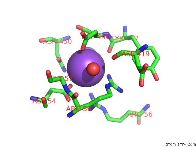
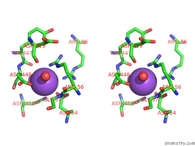
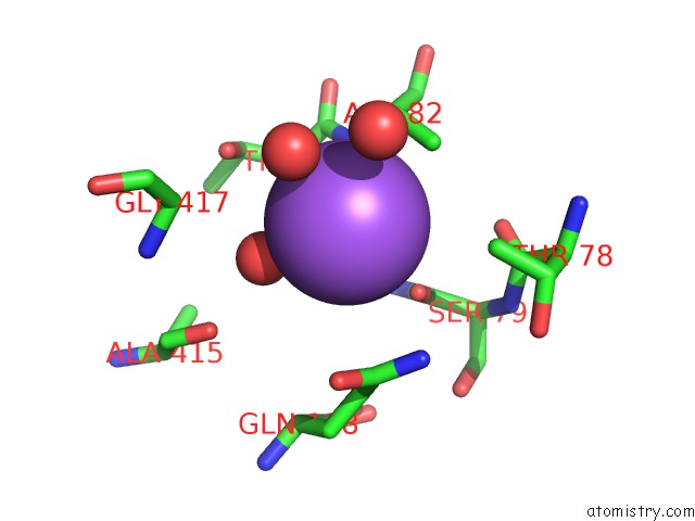
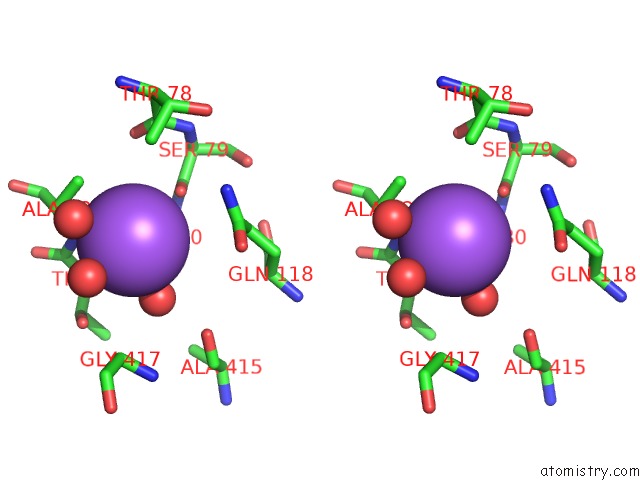
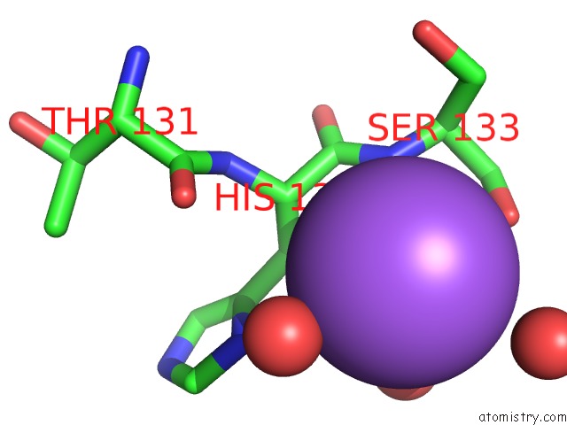
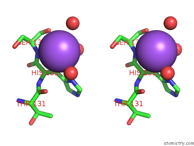
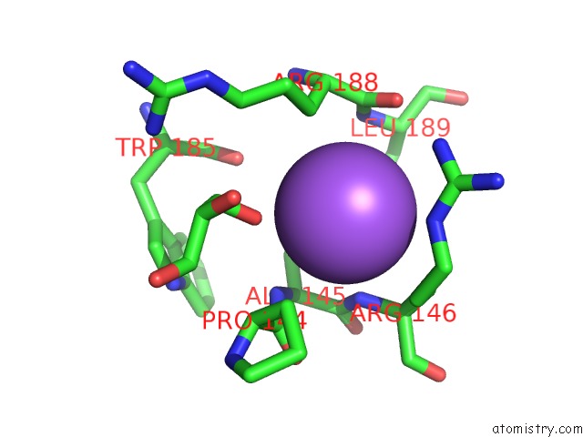
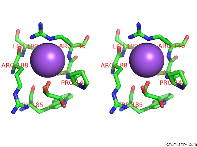
Sodium binding site 1 out of 37 in 3hww
Go back to
Sodium binding site 1 out
of 37 in the Crystal Structure of Menaquinone Synthesis Protein Mend From E. Coli in Complex with Oxoglutarate
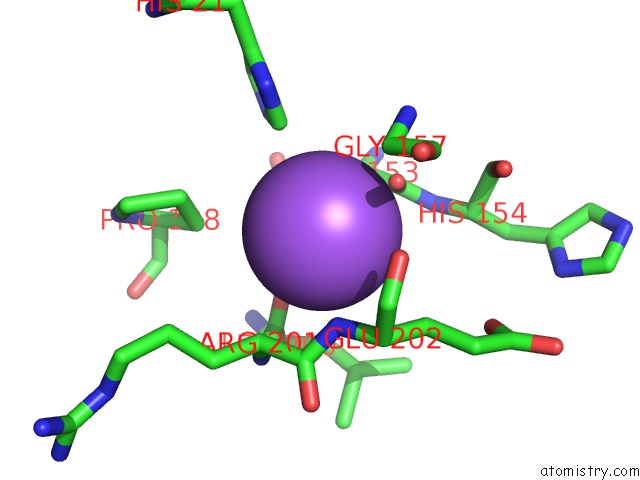
Mono view
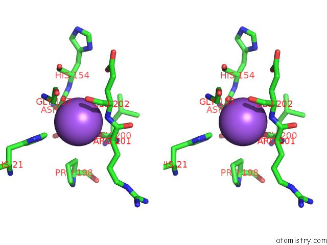
Stereo pair view
Mono view
Stereo pair view
A full contact list of Sodium with other atoms in the Na binding
site number 1 of Crystal Structure of Menaquinone Synthesis Protein Mend From E. Coli in Complex with Oxoglutarate within 5.0Å range:
|
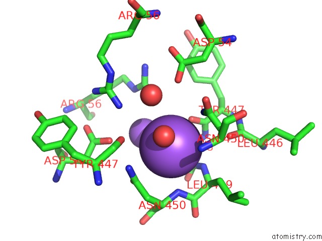
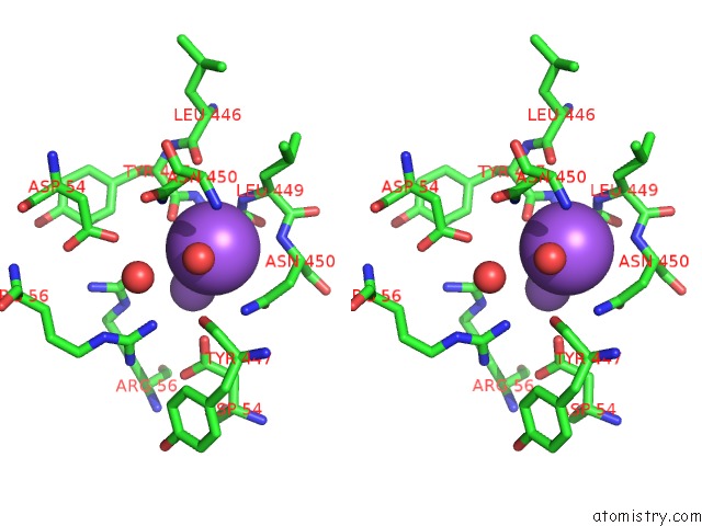
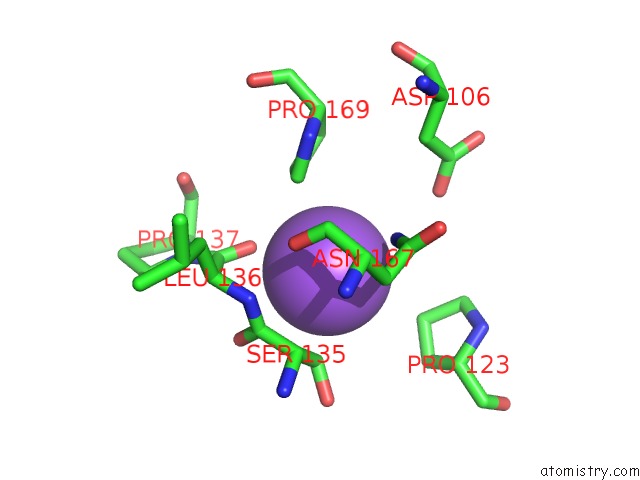
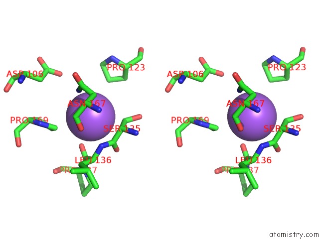
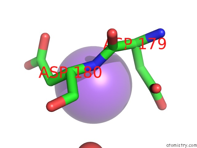
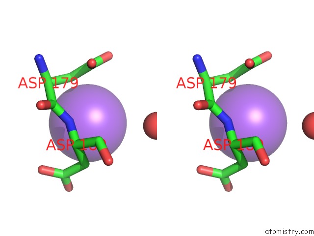
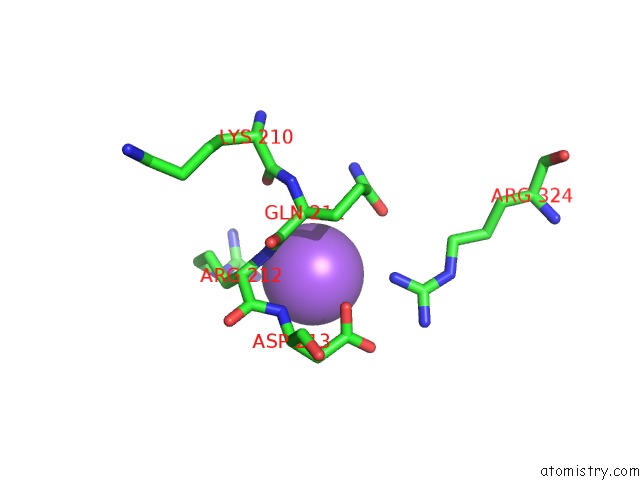
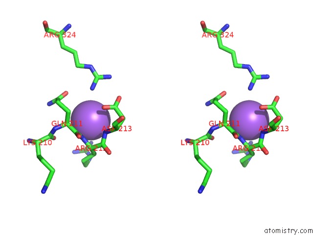
Sodium binding site 2 out of 37 in 3hww
Go back to
Sodium binding site 2 out
of 37 in the Crystal Structure of Menaquinone Synthesis Protein Mend From E. Coli in Complex with Oxoglutarate
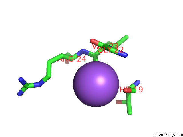
Mono view
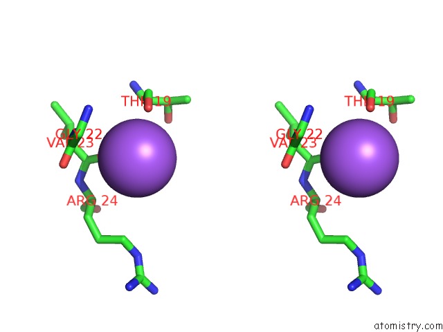
Stereo pair view
Mono view
Stereo pair view
A full contact list of Sodium with other atoms in the Na binding
site number 2 of Crystal Structure of Menaquinone Synthesis Protein Mend From E. Coli in Complex with Oxoglutarate within 5.0Å range:
|
Sodium binding site 3 out of 37 in 3hww
Go back to
Sodium binding site 3 out
of 37 in the Crystal Structure of Menaquinone Synthesis Protein Mend From E. Coli in Complex with Oxoglutarate
Mono view
Stereo pair view
Mono view
Stereo pair view
A full contact list of Sodium with other atoms in the Na binding
site number 3 of Crystal Structure of Menaquinone Synthesis Protein Mend From E. Coli in Complex with Oxoglutarate within 5.0Å range:
|
Sodium binding site 4 out of 37 in 3hww
Go back to
Sodium binding site 4 out
of 37 in the Crystal Structure of Menaquinone Synthesis Protein Mend From E. Coli in Complex with Oxoglutarate
Mono view
Stereo pair view
Mono view
Stereo pair view
A full contact list of Sodium with other atoms in the Na binding
site number 4 of Crystal Structure of Menaquinone Synthesis Protein Mend From E. Coli in Complex with Oxoglutarate within 5.0Å range:
|
Sodium binding site 5 out of 37 in 3hww
Go back to
Sodium binding site 5 out
of 37 in the Crystal Structure of Menaquinone Synthesis Protein Mend From E. Coli in Complex with Oxoglutarate
Mono view
Stereo pair view
Mono view
Stereo pair view
A full contact list of Sodium with other atoms in the Na binding
site number 5 of Crystal Structure of Menaquinone Synthesis Protein Mend From E. Coli in Complex with Oxoglutarate within 5.0Å range:
|
Sodium binding site 6 out of 37 in 3hww
Go back to
Sodium binding site 6 out
of 37 in the Crystal Structure of Menaquinone Synthesis Protein Mend From E. Coli in Complex with Oxoglutarate
Mono view
Stereo pair view
Mono view
Stereo pair view
A full contact list of Sodium with other atoms in the Na binding
site number 6 of Crystal Structure of Menaquinone Synthesis Protein Mend From E. Coli in Complex with Oxoglutarate within 5.0Å range:
|
Sodium binding site 7 out of 37 in 3hww
Go back to
Sodium binding site 7 out
of 37 in the Crystal Structure of Menaquinone Synthesis Protein Mend From E. Coli in Complex with Oxoglutarate
Mono view
Stereo pair view
Mono view
Stereo pair view
A full contact list of Sodium with other atoms in the Na binding
site number 7 of Crystal Structure of Menaquinone Synthesis Protein Mend From E. Coli in Complex with Oxoglutarate within 5.0Å range:
|
Sodium binding site 8 out of 37 in 3hww
Go back to
Sodium binding site 8 out
of 37 in the Crystal Structure of Menaquinone Synthesis Protein Mend From E. Coli in Complex with Oxoglutarate
Mono view
Stereo pair view
Mono view
Stereo pair view
A full contact list of Sodium with other atoms in the Na binding
site number 8 of Crystal Structure of Menaquinone Synthesis Protein Mend From E. Coli in Complex with Oxoglutarate within 5.0Å range:
|
Sodium binding site 9 out of 37 in 3hww
Go back to
Sodium binding site 9 out
of 37 in the Crystal Structure of Menaquinone Synthesis Protein Mend From E. Coli in Complex with Oxoglutarate
Mono view
Stereo pair view
Mono view
Stereo pair view
A full contact list of Sodium with other atoms in the Na binding
site number 9 of Crystal Structure of Menaquinone Synthesis Protein Mend From E. Coli in Complex with Oxoglutarate within 5.0Å range:
|
Sodium binding site 10 out of 37 in 3hww
Go back to
Sodium binding site 10 out
of 37 in the Crystal Structure of Menaquinone Synthesis Protein Mend From E. Coli in Complex with Oxoglutarate
Mono view
Stereo pair view
Mono view
Stereo pair view
A full contact list of Sodium with other atoms in the Na binding
site number 10 of Crystal Structure of Menaquinone Synthesis Protein Mend From E. Coli in Complex with Oxoglutarate within 5.0Å range:
|
Reference:
A.Priyadarshi,
E.E.Kim,
K.Y.Hwang.
Structural and Functional Analysis of Vitamin K2 Synthesis Protein Mend. Biochem.Biophys.Res.Commun. V. 388 748 2009.
ISSN: ISSN 0006-291X
PubMed: 19703421
DOI: 10.1016/J.BBRC.2009.08.093
Page generated: Mon Oct 7 10:29:06 2024
ISSN: ISSN 0006-291X
PubMed: 19703421
DOI: 10.1016/J.BBRC.2009.08.093
Last articles
Zn in 9MJ5Zn in 9HNW
Zn in 9G0L
Zn in 9FNE
Zn in 9DZN
Zn in 9E0I
Zn in 9D32
Zn in 9DAK
Zn in 8ZXC
Zn in 8ZUF