Sodium »
PDB 3h0n-3hwd »
3hij »
Sodium in PDB 3hij: Crystal Structure of Dihydrodipicolinate Synthase From Bacillus Anthracis in Complex with Its Substrate, Pyruvate
Protein crystallography data
The structure of Crystal Structure of Dihydrodipicolinate Synthase From Bacillus Anthracis in Complex with Its Substrate, Pyruvate, PDB code: 3hij
was solved by
J.E.Voss,
S.W.Scally,
R.C.J.Dobson,
M.A.Perugini,
with X-Ray Crystallography technique. A brief refinement statistics is given in the table below:
| Resolution Low / High (Å) | 37.27 / 2.15 |
| Space group | P 21 21 21 |
| Cell size a, b, c (Å), α, β, γ (°) | 84.484, 124.617, 130.979, 90.00, 90.00, 90.00 |
| R / Rfree (%) | 15.3 / 21 |
Sodium Binding Sites:
The binding sites of Sodium atom in the Crystal Structure of Dihydrodipicolinate Synthase From Bacillus Anthracis in Complex with Its Substrate, Pyruvate
(pdb code 3hij). This binding sites where shown within
5.0 Angstroms radius around Sodium atom.
In total 4 binding sites of Sodium where determined in the Crystal Structure of Dihydrodipicolinate Synthase From Bacillus Anthracis in Complex with Its Substrate, Pyruvate, PDB code: 3hij:
Jump to Sodium binding site number: 1; 2; 3; 4;
In total 4 binding sites of Sodium where determined in the Crystal Structure of Dihydrodipicolinate Synthase From Bacillus Anthracis in Complex with Its Substrate, Pyruvate, PDB code: 3hij:
Jump to Sodium binding site number: 1; 2; 3; 4;
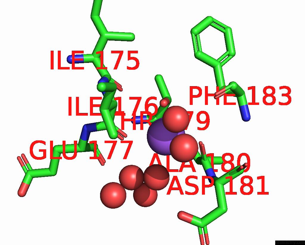
Sodium binding site 1 out of 4 in 3hij
Go back to
Sodium binding site 1 out
of 4 in the Crystal Structure of Dihydrodipicolinate Synthase From Bacillus Anthracis in Complex with Its Substrate, Pyruvate
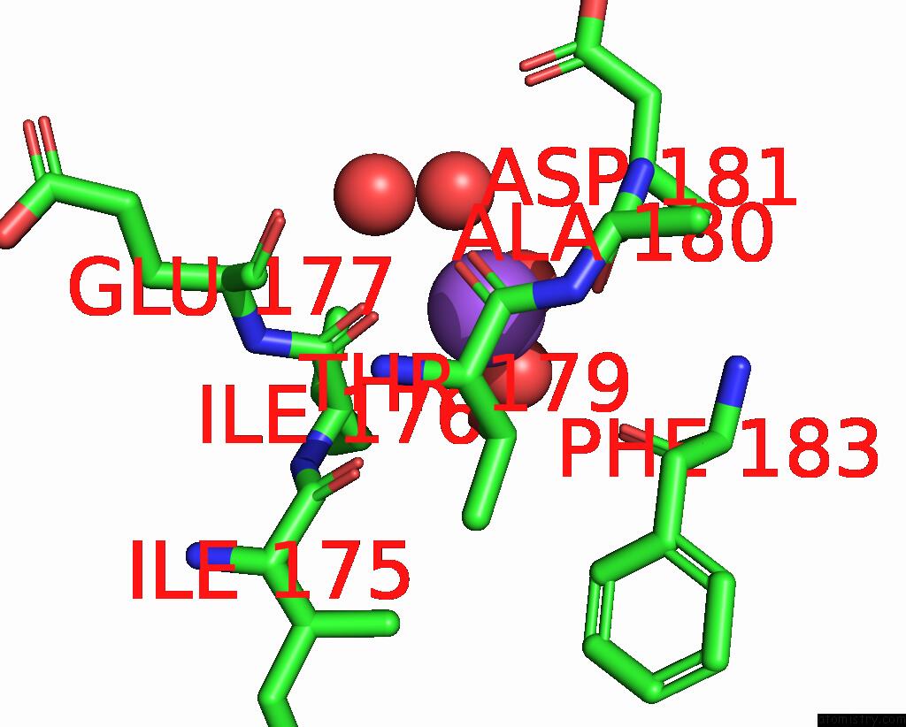
Mono view

Stereo pair view
Mono view
Stereo pair view
A full contact list of Sodium with other atoms in the Na binding
site number 1 of Crystal Structure of Dihydrodipicolinate Synthase From Bacillus Anthracis in Complex with Its Substrate, Pyruvate within 5.0Å range:
|
Sodium binding site 2 out of 4 in 3hij
Go back to
Sodium binding site 2 out
of 4 in the Crystal Structure of Dihydrodipicolinate Synthase From Bacillus Anthracis in Complex with Its Substrate, Pyruvate
Mono view

Stereo pair view
Mono view
Stereo pair view
A full contact list of Sodium with other atoms in the Na binding
site number 2 of Crystal Structure of Dihydrodipicolinate Synthase From Bacillus Anthracis in Complex with Its Substrate, Pyruvate within 5.0Å range:
|
Sodium binding site 3 out of 4 in 3hij
Go back to
Sodium binding site 3 out
of 4 in the Crystal Structure of Dihydrodipicolinate Synthase From Bacillus Anthracis in Complex with Its Substrate, Pyruvate

Mono view

Stereo pair view
Mono view
Stereo pair view
A full contact list of Sodium with other atoms in the Na binding
site number 3 of Crystal Structure of Dihydrodipicolinate Synthase From Bacillus Anthracis in Complex with Its Substrate, Pyruvate within 5.0Å range:
|
Sodium binding site 4 out of 4 in 3hij
Go back to
Sodium binding site 4 out
of 4 in the Crystal Structure of Dihydrodipicolinate Synthase From Bacillus Anthracis in Complex with Its Substrate, Pyruvate

Mono view

Stereo pair view
Mono view
Stereo pair view
A full contact list of Sodium with other atoms in the Na binding
site number 4 of Crystal Structure of Dihydrodipicolinate Synthase From Bacillus Anthracis in Complex with Its Substrate, Pyruvate within 5.0Å range:
|
Reference:
J.E.Voss,
S.W.Scally,
N.L.Taylor,
S.C.Atkinson,
M.D.Griffin,
C.A.Hutton,
M.W.Parker,
M.R.Alderton,
J.A.Gerrard,
R.C.Dobson,
C.Dogovski,
M.A.Perugini.
Substrate-Mediated Stabilization of A Tetrameric Drug Target Reveals Achilles Heel in Anthrax. J.Biol.Chem. V. 285 5188 2010.
ISSN: ISSN 0021-9258
PubMed: 19948665
DOI: 10.1074/JBC.M109.038166
Page generated: Sun Aug 17 15:19:27 2025
ISSN: ISSN 0021-9258
PubMed: 19948665
DOI: 10.1074/JBC.M109.038166
Last articles
Na in 4XKWNa in 4XL9
Na in 4XKY
Na in 4XK2
Na in 4XKV
Na in 4XKR
Na in 4XHS
Na in 4XJI
Na in 4XJH
Na in 4XJF