Sodium »
PDB 3gdx-3h09 »
3goa »
Sodium in PDB 3goa: Crystal Structure of the Salmonella Typhimurium Fada 3-Ketoacyl-Coa Thiolase
Enzymatic activity of Crystal Structure of the Salmonella Typhimurium Fada 3-Ketoacyl-Coa Thiolase
All present enzymatic activity of Crystal Structure of the Salmonella Typhimurium Fada 3-Ketoacyl-Coa Thiolase:
2.3.1.16;
2.3.1.16;
Protein crystallography data
The structure of Crystal Structure of the Salmonella Typhimurium Fada 3-Ketoacyl-Coa Thiolase, PDB code: 3goa
was solved by
S.M.Anderson,
T.Skarina,
O.Onopriyenko,
Z.Wawrzak,
L.Papazisi,
A.Savchenko,
W.F.Anderson,
Center For Structural Genomics Ofinfectious Diseases (Csgid),
with X-Ray Crystallography technique. A brief refinement statistics is given in the table below:
| Resolution Low / High (Å) | 30.00 / 1.70 |
| Space group | P 1 21 1 |
| Cell size a, b, c (Å), α, β, γ (°) | 73.000, 64.500, 74.400, 90.00, 104.50, 90.00 |
| R / Rfree (%) | 14.2 / 19.2 |
Other elements in 3goa:
The structure of Crystal Structure of the Salmonella Typhimurium Fada 3-Ketoacyl-Coa Thiolase also contains other interesting chemical elements:
| Calcium | (Ca) | 2 atoms |
| Chlorine | (Cl) | 2 atoms |
Sodium Binding Sites:
The binding sites of Sodium atom in the Crystal Structure of the Salmonella Typhimurium Fada 3-Ketoacyl-Coa Thiolase
(pdb code 3goa). This binding sites where shown within
5.0 Angstroms radius around Sodium atom.
In total only one binding site of Sodium was determined in the Crystal Structure of the Salmonella Typhimurium Fada 3-Ketoacyl-Coa Thiolase, PDB code: 3goa:
In total only one binding site of Sodium was determined in the Crystal Structure of the Salmonella Typhimurium Fada 3-Ketoacyl-Coa Thiolase, PDB code: 3goa:
Sodium binding site 1 out of 1 in 3goa
Go back to
Sodium binding site 1 out
of 1 in the Crystal Structure of the Salmonella Typhimurium Fada 3-Ketoacyl-Coa Thiolase
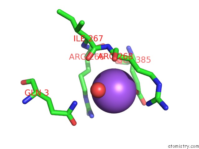
Mono view
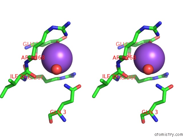
Stereo pair view
Mono view
Stereo pair view
A full contact list of Sodium with other atoms in the Na binding
site number 1 of Crystal Structure of the Salmonella Typhimurium Fada 3-Ketoacyl-Coa Thiolase within 5.0Å range:
|
Reference:
S.M.Anderson,
T.Skarina,
O.Onopriyenko,
Z.Wawrzak,
L.Papazisi,
A.Savchenko,
W.F.Anderson,
Center For Structural Genomics Ofinfectious Diseases (Csgid).
N/A N/A.
Page generated: Mon Oct 7 10:16:27 2024
Last articles
Cl in 5IJ7Cl in 5II3
Cl in 5IFM
Cl in 5IJ6
Cl in 5IHG
Cl in 5IHR
Cl in 5IGN
Cl in 5IH5
Cl in 5IG6
Cl in 5IFU