Sodium »
PDB 3e40-3ept »
3epg »
Sodium in PDB 3epg: Structure of Human Dna Polymerase Iota Complexed with N2-Ethylguanine
Enzymatic activity of Structure of Human Dna Polymerase Iota Complexed with N2-Ethylguanine
All present enzymatic activity of Structure of Human Dna Polymerase Iota Complexed with N2-Ethylguanine:
2.7.7.7;
2.7.7.7;
Protein crystallography data
The structure of Structure of Human Dna Polymerase Iota Complexed with N2-Ethylguanine, PDB code: 3epg
was solved by
M.G.Pence,
with X-Ray Crystallography technique. A brief refinement statistics is given in the table below:
| Resolution Low / High (Å) | 15.00 / 2.50 |
| Space group | P 65 2 2 |
| Cell size a, b, c (Å), α, β, γ (°) | 98.535, 98.535, 202.354, 90.00, 90.00, 120.00 |
| R / Rfree (%) | 23.4 / 28.3 |
Sodium Binding Sites:
The binding sites of Sodium atom in the Structure of Human Dna Polymerase Iota Complexed with N2-Ethylguanine
(pdb code 3epg). This binding sites where shown within
5.0 Angstroms radius around Sodium atom.
In total 4 binding sites of Sodium where determined in the Structure of Human Dna Polymerase Iota Complexed with N2-Ethylguanine, PDB code: 3epg:
Jump to Sodium binding site number: 1; 2; 3; 4;
In total 4 binding sites of Sodium where determined in the Structure of Human Dna Polymerase Iota Complexed with N2-Ethylguanine, PDB code: 3epg:
Jump to Sodium binding site number: 1; 2; 3; 4;
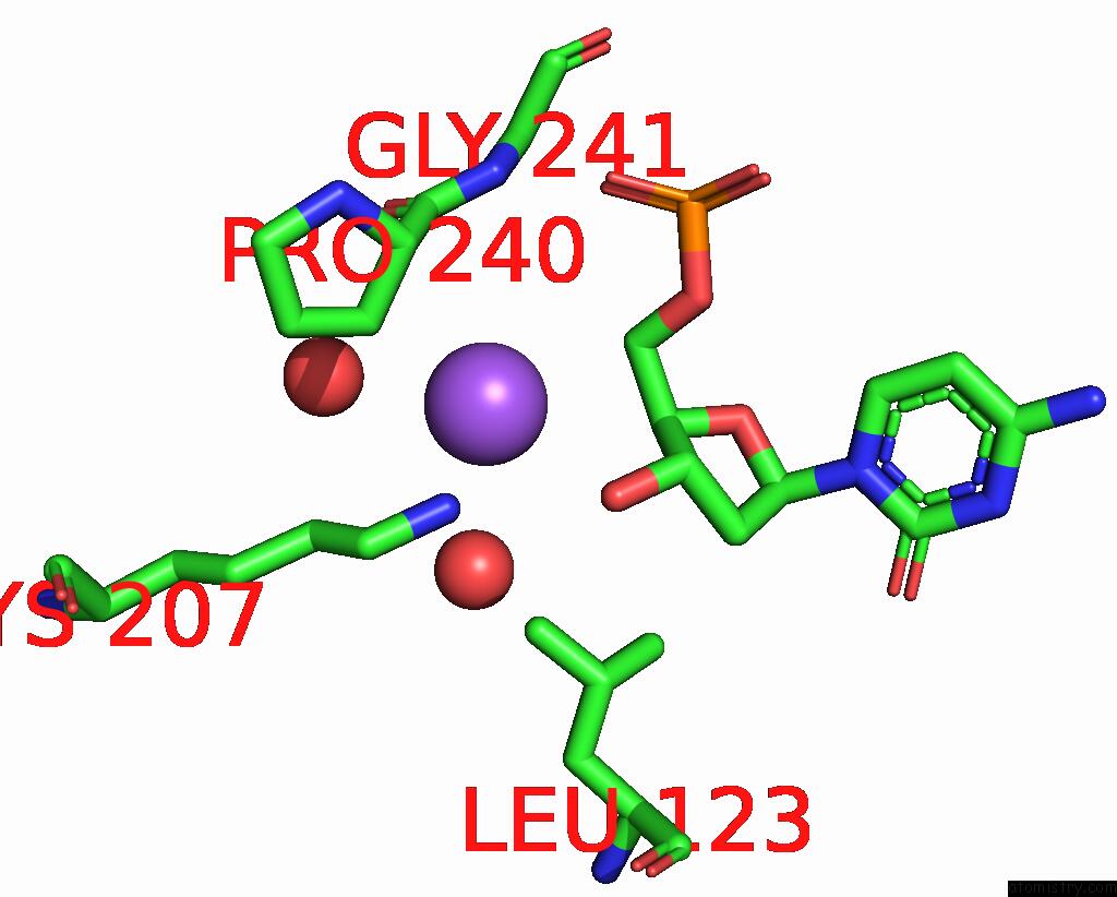
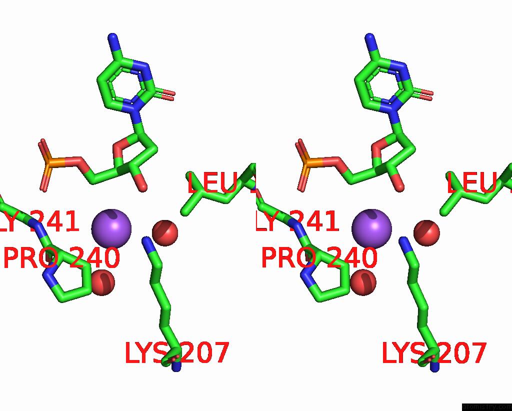
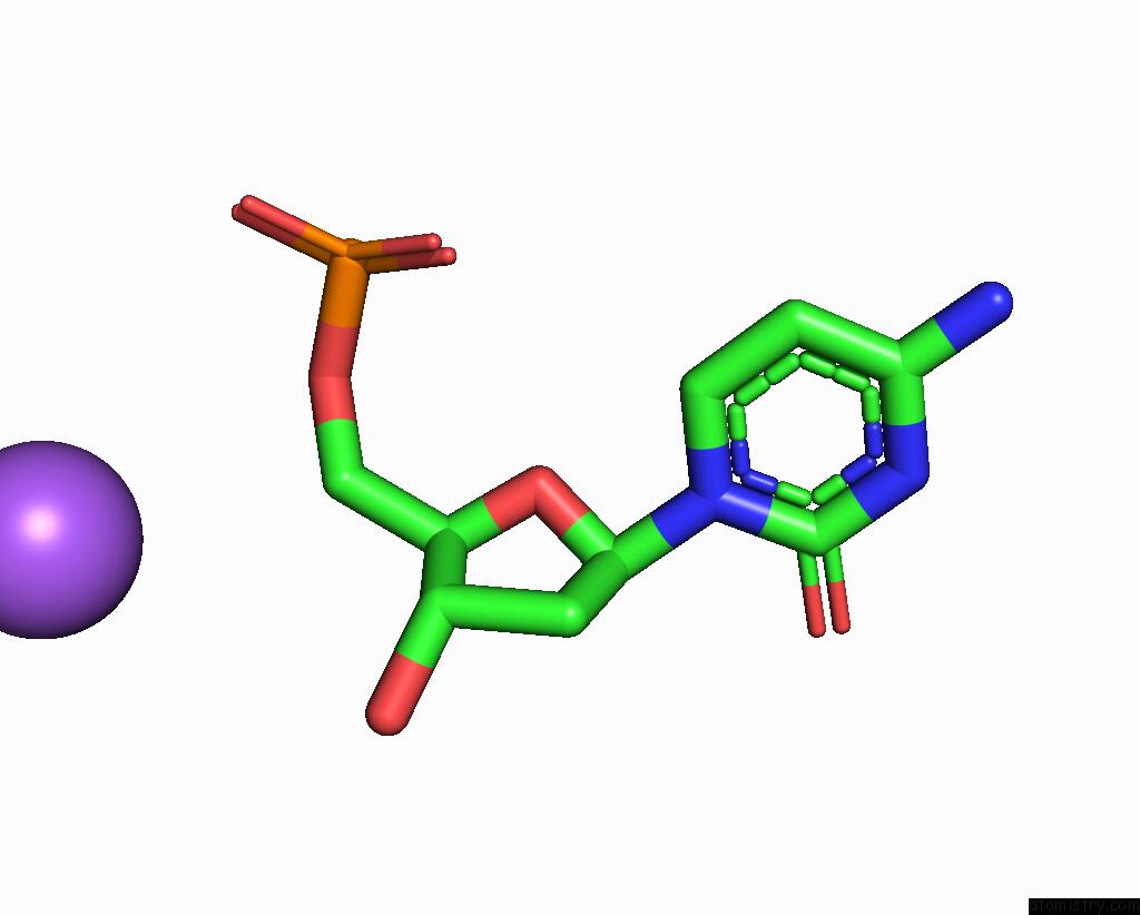
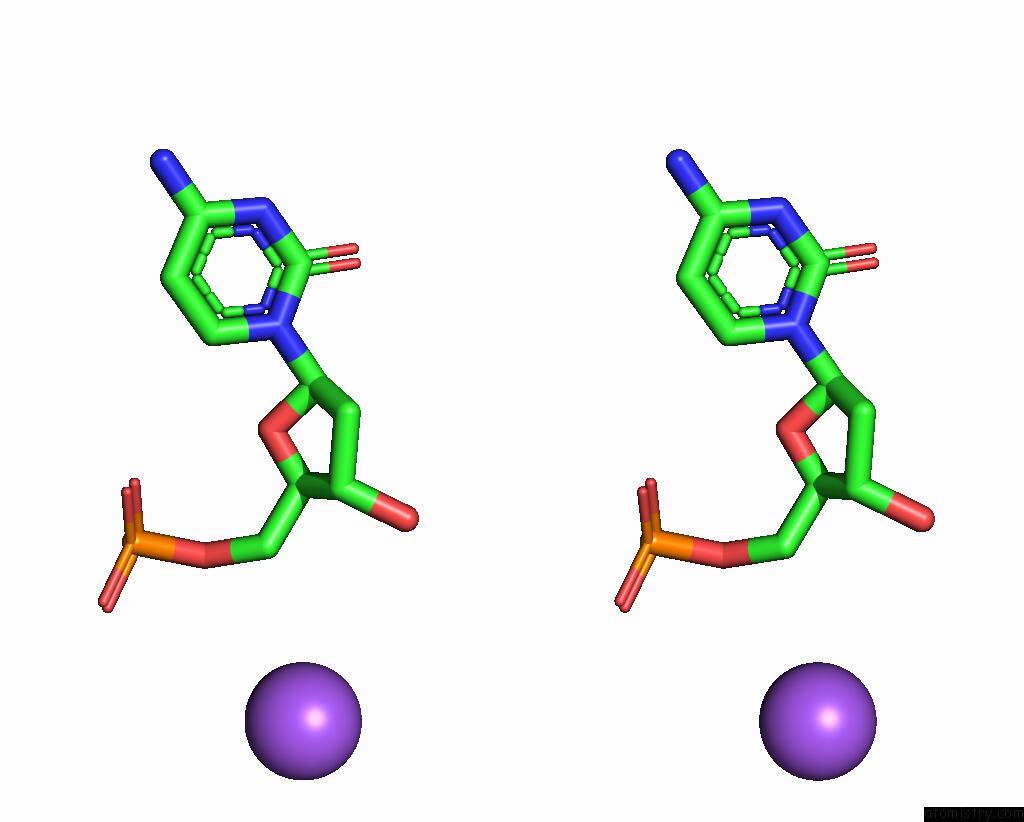
Sodium binding site 1 out of 4 in 3epg
Go back to
Sodium binding site 1 out
of 4 in the Structure of Human Dna Polymerase Iota Complexed with N2-Ethylguanine
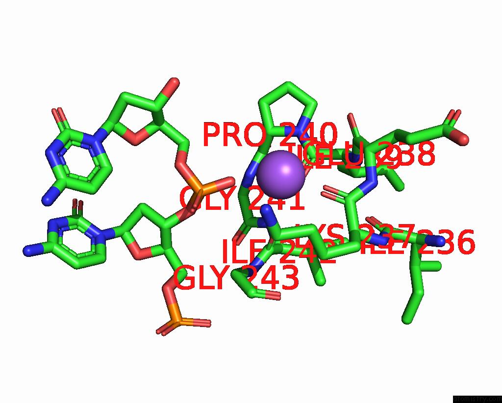
Mono view
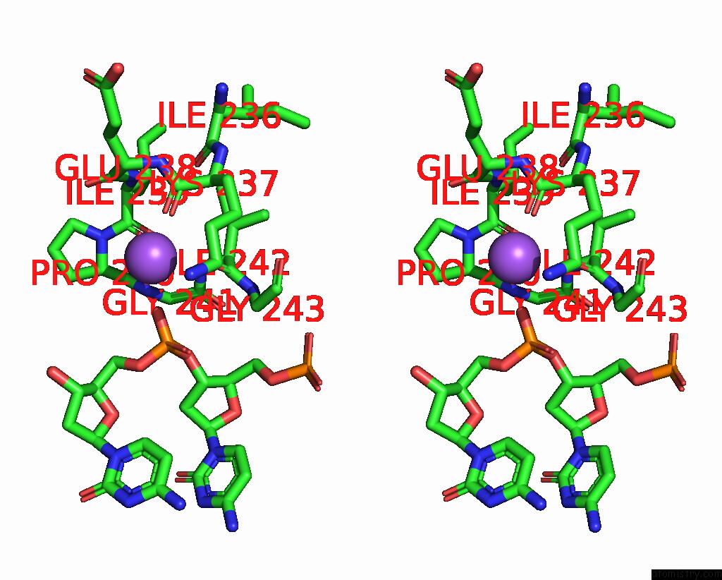
Stereo pair view
Mono view
Stereo pair view
A full contact list of Sodium with other atoms in the Na binding
site number 1 of Structure of Human Dna Polymerase Iota Complexed with N2-Ethylguanine within 5.0Å range:
|
Sodium binding site 2 out of 4 in 3epg
Go back to
Sodium binding site 2 out
of 4 in the Structure of Human Dna Polymerase Iota Complexed with N2-Ethylguanine
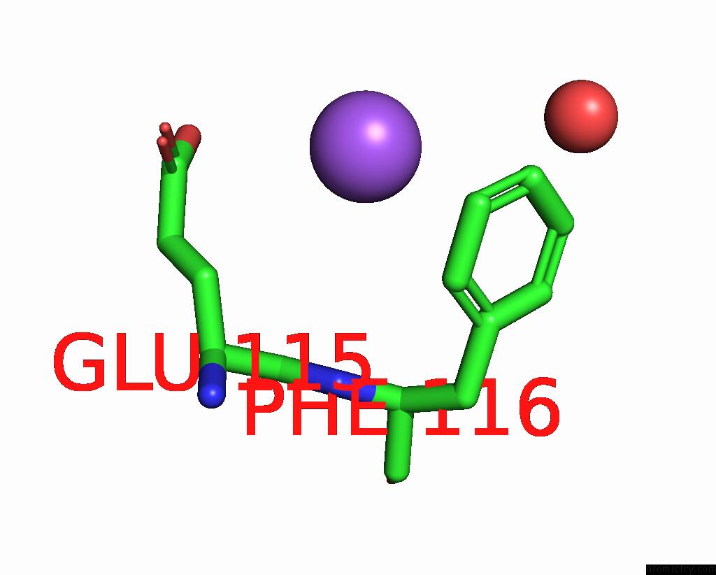
Mono view
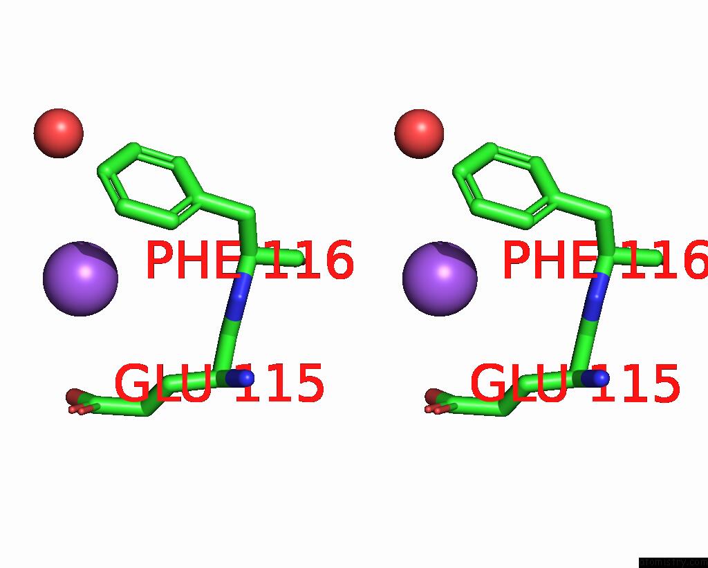
Stereo pair view
Mono view
Stereo pair view
A full contact list of Sodium with other atoms in the Na binding
site number 2 of Structure of Human Dna Polymerase Iota Complexed with N2-Ethylguanine within 5.0Å range:
|
Sodium binding site 3 out of 4 in 3epg
Go back to
Sodium binding site 3 out
of 4 in the Structure of Human Dna Polymerase Iota Complexed with N2-Ethylguanine
Mono view
Stereo pair view
Mono view
Stereo pair view
A full contact list of Sodium with other atoms in the Na binding
site number 3 of Structure of Human Dna Polymerase Iota Complexed with N2-Ethylguanine within 5.0Å range:
|
Sodium binding site 4 out of 4 in 3epg
Go back to
Sodium binding site 4 out
of 4 in the Structure of Human Dna Polymerase Iota Complexed with N2-Ethylguanine
Mono view
Stereo pair view
Mono view
Stereo pair view
A full contact list of Sodium with other atoms in the Na binding
site number 4 of Structure of Human Dna Polymerase Iota Complexed with N2-Ethylguanine within 5.0Å range:
|
Reference:
M.G.Pence,
P.Blans,
C.N.Zink,
T.Hollis,
J.C.Fishbein,
F.W.Perrino.
Lesion Bypass of N2-Ethylguanine By Human Dna Polymerase Iota. J.Biol.Chem. V. 284 1732 2009.
ISSN: ISSN 0021-9258
PubMed: 18984581
DOI: 10.1074/JBC.M807296200
Page generated: Mon Oct 7 08:52:31 2024
ISSN: ISSN 0021-9258
PubMed: 18984581
DOI: 10.1074/JBC.M807296200
Last articles
F in 7NVMF in 7NW2
F in 7NW0
F in 7NVL
F in 7NVX
F in 7NVV
F in 7NVO
F in 7NTH
F in 7NTI
F in 7NPC