Sodium »
PDB 3e40-3ept »
3efp »
Sodium in PDB 3efp: Crystal Structure of the Escherichia Coli Twin Arginine Leader Peptide Binding Protein Dmsd in A Monomeric Form
Protein crystallography data
The structure of Crystal Structure of the Escherichia Coli Twin Arginine Leader Peptide Binding Protein Dmsd in A Monomeric Form, PDB code: 3efp
was solved by
C.M.Stevens,
M.Paetzel,
with X-Ray Crystallography technique. A brief refinement statistics is given in the table below:
| Resolution Low / High (Å) | 30.75 / 2.01 |
| Space group | P 31 2 1 |
| Cell size a, b, c (Å), α, β, γ (°) | 128.020, 128.020, 78.723, 90.00, 90.00, 120.00 |
| R / Rfree (%) | 17.8 / 21.3 |
Other elements in 3efp:
The structure of Crystal Structure of the Escherichia Coli Twin Arginine Leader Peptide Binding Protein Dmsd in A Monomeric Form also contains other interesting chemical elements:
| Chlorine | (Cl) | 6 atoms |
Sodium Binding Sites:
The binding sites of Sodium atom in the Crystal Structure of the Escherichia Coli Twin Arginine Leader Peptide Binding Protein Dmsd in A Monomeric Form
(pdb code 3efp). This binding sites where shown within
5.0 Angstroms radius around Sodium atom.
In total only one binding site of Sodium was determined in the Crystal Structure of the Escherichia Coli Twin Arginine Leader Peptide Binding Protein Dmsd in A Monomeric Form, PDB code: 3efp:
In total only one binding site of Sodium was determined in the Crystal Structure of the Escherichia Coli Twin Arginine Leader Peptide Binding Protein Dmsd in A Monomeric Form, PDB code: 3efp:
Sodium binding site 1 out of 1 in 3efp
Go back to
Sodium binding site 1 out
of 1 in the Crystal Structure of the Escherichia Coli Twin Arginine Leader Peptide Binding Protein Dmsd in A Monomeric Form
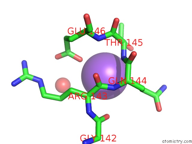
Mono view
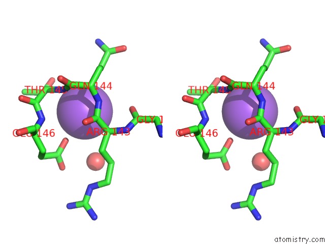
Stereo pair view
Mono view
Stereo pair view
A full contact list of Sodium with other atoms in the Na binding
site number 1 of Crystal Structure of the Escherichia Coli Twin Arginine Leader Peptide Binding Protein Dmsd in A Monomeric Form within 5.0Å range:
|
Reference:
C.M.Stevens,
T.M.Winstone,
R.J.Turner,
M.Paetzel.
Structural Analysis of A Monomeric Form of the Twin-Arginine Leader Peptide Binding Chaperone Escherichia Coli Dmsd. J.Mol.Biol. V. 389 124 2009.
ISSN: ISSN 0022-2836
PubMed: 19361518
DOI: 10.1016/J.JMB.2009.03.069
Page generated: Mon Oct 7 08:51:00 2024
ISSN: ISSN 0022-2836
PubMed: 19361518
DOI: 10.1016/J.JMB.2009.03.069
Last articles
Zn in 9MJ5Zn in 9HNW
Zn in 9G0L
Zn in 9FNE
Zn in 9DZN
Zn in 9E0I
Zn in 9D32
Zn in 9DAK
Zn in 8ZXC
Zn in 8ZUF