Sodium »
PDB 3cq8-3dfh »
3ddr »
Sodium in PDB 3ddr: Structure of the Serratia Marcescens Hemophore Receptor Hasr-ILE671GLY Mutant in Complex with Its Hemophore Hasa and Heme
Protein crystallography data
The structure of Structure of the Serratia Marcescens Hemophore Receptor Hasr-ILE671GLY Mutant in Complex with Its Hemophore Hasa and Heme, PDB code: 3ddr
was solved by
S.Krieg,
K.Diederichs,
with X-Ray Crystallography technique. A brief refinement statistics is given in the table below:
| Resolution Low / High (Å) | 39.25 / 2.80 |
| Space group | F 2 2 2 |
| Cell size a, b, c (Å), α, β, γ (°) | 157.410, 162.720, 597.010, 90.00, 90.00, 90.00 |
| R / Rfree (%) | 22.6 / 26.2 |
Other elements in 3ddr:
The structure of Structure of the Serratia Marcescens Hemophore Receptor Hasr-ILE671GLY Mutant in Complex with Its Hemophore Hasa and Heme also contains other interesting chemical elements:
| Iron | (Fe) | 2 atoms |
Sodium Binding Sites:
The binding sites of Sodium atom in the Structure of the Serratia Marcescens Hemophore Receptor Hasr-ILE671GLY Mutant in Complex with Its Hemophore Hasa and Heme
(pdb code 3ddr). This binding sites where shown within
5.0 Angstroms radius around Sodium atom.
In total 2 binding sites of Sodium where determined in the Structure of the Serratia Marcescens Hemophore Receptor Hasr-ILE671GLY Mutant in Complex with Its Hemophore Hasa and Heme, PDB code: 3ddr:
Jump to Sodium binding site number: 1; 2;
In total 2 binding sites of Sodium where determined in the Structure of the Serratia Marcescens Hemophore Receptor Hasr-ILE671GLY Mutant in Complex with Its Hemophore Hasa and Heme, PDB code: 3ddr:
Jump to Sodium binding site number: 1; 2;
Sodium binding site 1 out of 2 in 3ddr
Go back to
Sodium binding site 1 out
of 2 in the Structure of the Serratia Marcescens Hemophore Receptor Hasr-ILE671GLY Mutant in Complex with Its Hemophore Hasa and Heme
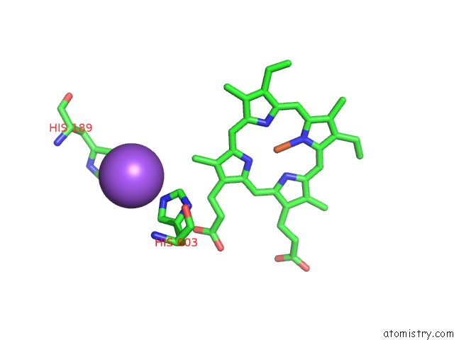
Mono view
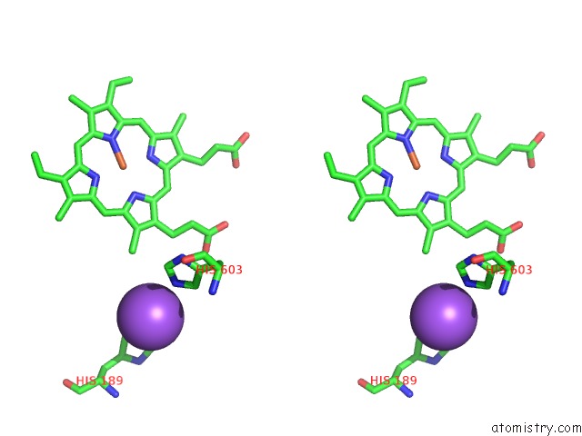
Stereo pair view
Mono view
Stereo pair view
A full contact list of Sodium with other atoms in the Na binding
site number 1 of Structure of the Serratia Marcescens Hemophore Receptor Hasr-ILE671GLY Mutant in Complex with Its Hemophore Hasa and Heme within 5.0Å range:
|
Sodium binding site 2 out of 2 in 3ddr
Go back to
Sodium binding site 2 out
of 2 in the Structure of the Serratia Marcescens Hemophore Receptor Hasr-ILE671GLY Mutant in Complex with Its Hemophore Hasa and Heme
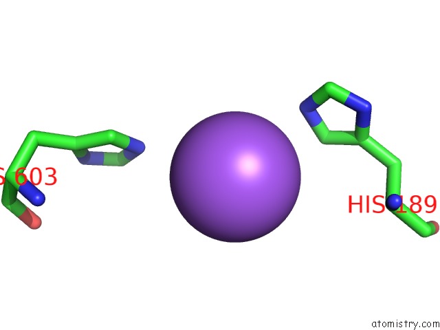
Mono view
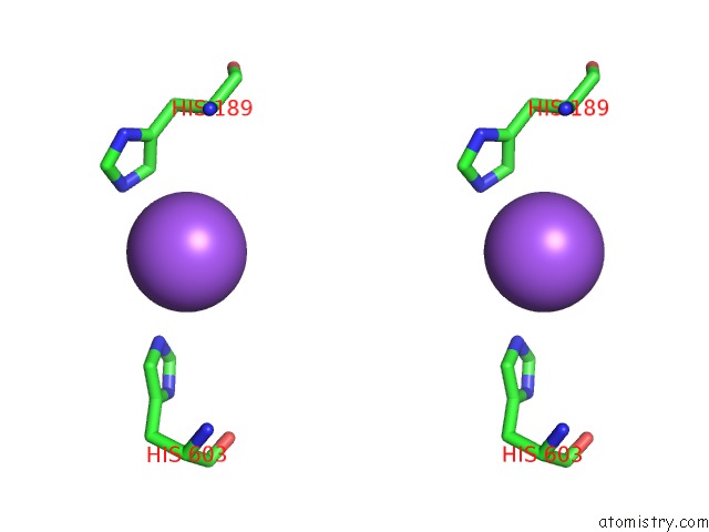
Stereo pair view
Mono view
Stereo pair view
A full contact list of Sodium with other atoms in the Na binding
site number 2 of Structure of the Serratia Marcescens Hemophore Receptor Hasr-ILE671GLY Mutant in Complex with Its Hemophore Hasa and Heme within 5.0Å range:
|
Reference:
S.Krieg,
F.Huche,
K.Diederichs,
N.Izadi-Pruneyre,
A.Lecroisey,
C.Wandersman,
P.Delepelaire,
W.Welte.
Heme Uptake Across the Outer Membrane As Revealed By Crystal Structures of the Receptor-Hemophore Complex. Proc.Natl.Acad.Sci.Usa V. 106 1045 2009.
ISSN: ISSN 0027-8424
PubMed: 19144921
DOI: 10.1073/PNAS.0809406106
Page generated: Mon Oct 7 08:12:29 2024
ISSN: ISSN 0027-8424
PubMed: 19144921
DOI: 10.1073/PNAS.0809406106
Last articles
Cl in 5QACCl in 5QAD
Cl in 5QAA
Cl in 5QAB
Cl in 5QA9
Cl in 5QA7
Cl in 5QA6
Cl in 5QA8
Cl in 5QA5
Cl in 5QA4