Sodium »
PDB 3cq8-3dfh »
3d76 »
Sodium in PDB 3d76: Crystal Structure of A Pheromone Binding Protein Mutant D35N, From Apis Mellifera, Soaked at pH 7.0
Protein crystallography data
The structure of Crystal Structure of A Pheromone Binding Protein Mutant D35N, From Apis Mellifera, Soaked at pH 7.0, PDB code: 3d76
was solved by
M.E.Pesenti,
S.Spinelli,
V.Bezirard,
L.Briand,
J.C.Pernollet,
M.Tegoni,
C.Cambillau,
with X-Ray Crystallography technique. A brief refinement statistics is given in the table below:
| Resolution Low / High (Å) | 15.00 / 1.90 |
| Space group | C 2 2 21 |
| Cell size a, b, c (Å), α, β, γ (°) | 78.991, 84.196, 47.558, 90.00, 90.00, 90.00 |
| R / Rfree (%) | 19 / 24.1 |
Sodium Binding Sites:
The binding sites of Sodium atom in the Crystal Structure of A Pheromone Binding Protein Mutant D35N, From Apis Mellifera, Soaked at pH 7.0
(pdb code 3d76). This binding sites where shown within
5.0 Angstroms radius around Sodium atom.
In total only one binding site of Sodium was determined in the Crystal Structure of A Pheromone Binding Protein Mutant D35N, From Apis Mellifera, Soaked at pH 7.0, PDB code: 3d76:
In total only one binding site of Sodium was determined in the Crystal Structure of A Pheromone Binding Protein Mutant D35N, From Apis Mellifera, Soaked at pH 7.0, PDB code: 3d76:
Sodium binding site 1 out of 1 in 3d76
Go back to
Sodium binding site 1 out
of 1 in the Crystal Structure of A Pheromone Binding Protein Mutant D35N, From Apis Mellifera, Soaked at pH 7.0
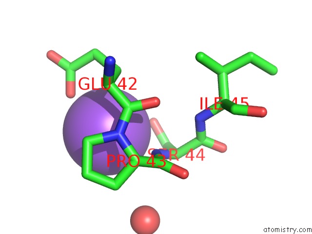
Mono view
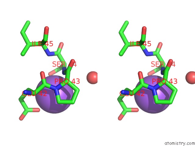
Stereo pair view
Mono view
Stereo pair view
A full contact list of Sodium with other atoms in the Na binding
site number 1 of Crystal Structure of A Pheromone Binding Protein Mutant D35N, From Apis Mellifera, Soaked at pH 7.0 within 5.0Å range:
|
Reference:
M.E.Pesenti,
S.Spinelli,
V.Bezirard,
L.Briand,
J.C.Pernollet,
V.Campanacci,
M.Tegoni,
C.Cambillau.
Queen Bee Pheromone Binding Protein pH-Induced Domain Swapping Favors Pheromone Release J.Mol.Biol. V. 390 981 2009.
ISSN: ISSN 0022-2836
PubMed: 19481550
DOI: 10.1016/J.JMB.2009.05.067
Page generated: Mon Oct 7 08:10:18 2024
ISSN: ISSN 0022-2836
PubMed: 19481550
DOI: 10.1016/J.JMB.2009.05.067
Last articles
Cl in 5FYSCl in 5FWV
Cl in 5FXT
Cl in 5FXS
Cl in 5FXR
Cl in 5FXQ
Cl in 5FWZ
Cl in 5FWE
Cl in 5FWU
Cl in 5FVZ