Sodium »
PDB 3c7o-3cpw »
3cmd »
Sodium in PDB 3cmd: Crystal Structure of Peptide Deformylase From Vre-E.Faecium
Enzymatic activity of Crystal Structure of Peptide Deformylase From Vre-E.Faecium
All present enzymatic activity of Crystal Structure of Peptide Deformylase From Vre-E.Faecium:
3.5.1.88;
3.5.1.88;
Protein crystallography data
The structure of Crystal Structure of Peptide Deformylase From Vre-E.Faecium, PDB code: 3cmd
was solved by
K.Y.Hwang,
K.H.Nam,
with X-Ray Crystallography technique. A brief refinement statistics is given in the table below:
| Resolution Low / High (Å) | 20.00 / 2.70 |
| Space group | P 64 2 2 |
| Cell size a, b, c (Å), α, β, γ (°) | 149.470, 149.470, 143.329, 90.00, 90.00, 120.00 |
| R / Rfree (%) | 20 / 22.9 |
Other elements in 3cmd:
The structure of Crystal Structure of Peptide Deformylase From Vre-E.Faecium also contains other interesting chemical elements:
| Iron | (Fe) | 2 atoms |
Sodium Binding Sites:
The binding sites of Sodium atom in the Crystal Structure of Peptide Deformylase From Vre-E.Faecium
(pdb code 3cmd). This binding sites where shown within
5.0 Angstroms radius around Sodium atom.
In total 8 binding sites of Sodium where determined in the Crystal Structure of Peptide Deformylase From Vre-E.Faecium, PDB code: 3cmd:
Jump to Sodium binding site number: 1; 2; 3; 4; 5; 6; 7; 8;
In total 8 binding sites of Sodium where determined in the Crystal Structure of Peptide Deformylase From Vre-E.Faecium, PDB code: 3cmd:
Jump to Sodium binding site number: 1; 2; 3; 4; 5; 6; 7; 8;
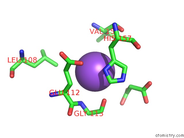
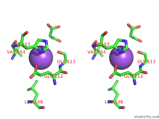
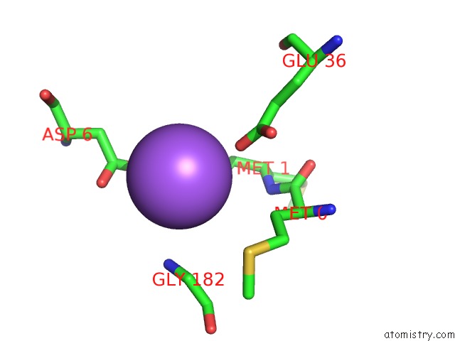
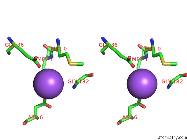
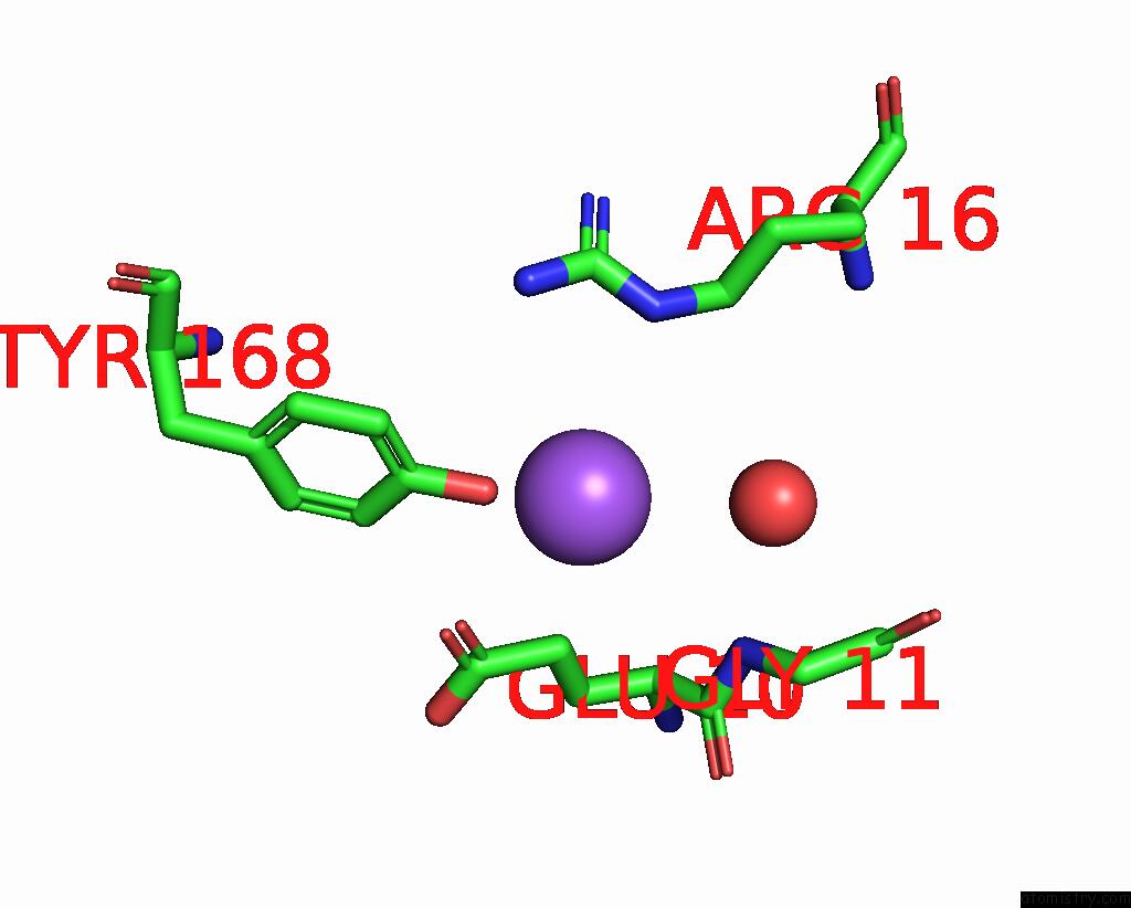
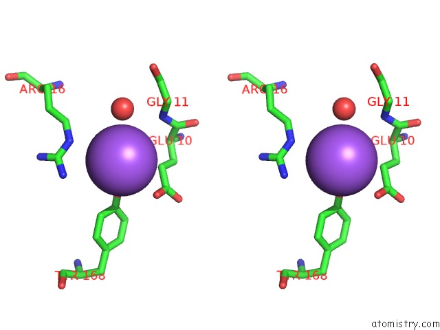
Sodium binding site 1 out of 8 in 3cmd
Go back to
Sodium binding site 1 out
of 8 in the Crystal Structure of Peptide Deformylase From Vre-E.Faecium
Mono view
Stereo pair view
Mono view
Stereo pair view
A full contact list of Sodium with other atoms in the Na binding
site number 1 of Crystal Structure of Peptide Deformylase From Vre-E.Faecium within 5.0Å range:
|
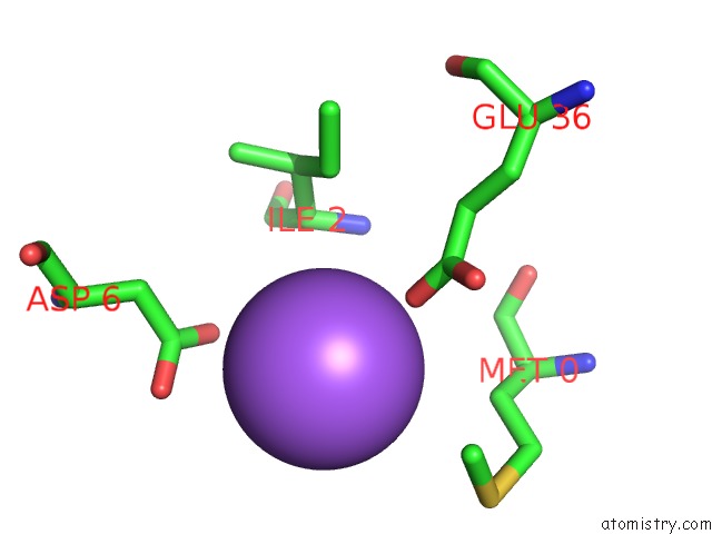
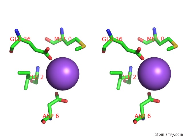
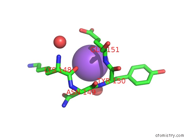
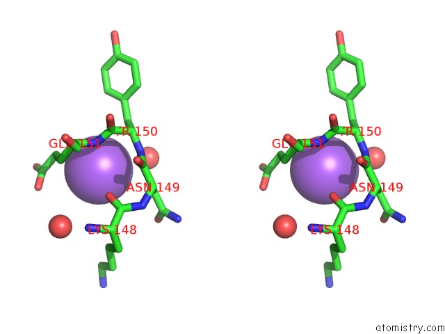
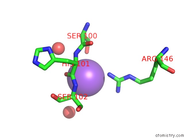
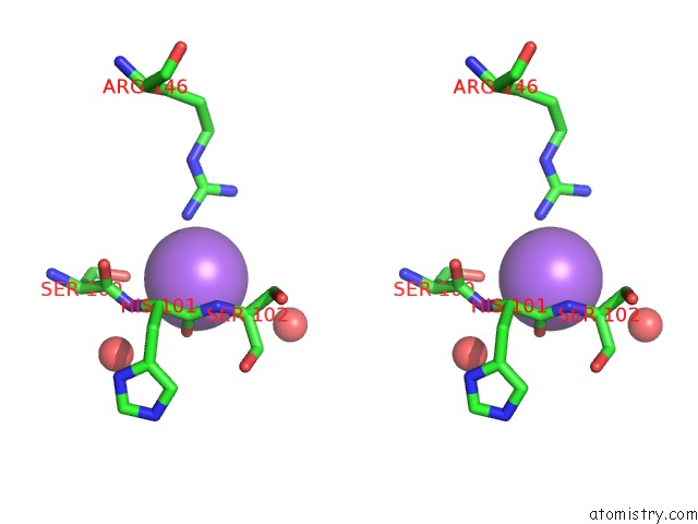
Sodium binding site 2 out of 8 in 3cmd
Go back to
Sodium binding site 2 out
of 8 in the Crystal Structure of Peptide Deformylase From Vre-E.Faecium
Mono view
Stereo pair view
Mono view
Stereo pair view
A full contact list of Sodium with other atoms in the Na binding
site number 2 of Crystal Structure of Peptide Deformylase From Vre-E.Faecium within 5.0Å range:
|
Sodium binding site 3 out of 8 in 3cmd
Go back to
Sodium binding site 3 out
of 8 in the Crystal Structure of Peptide Deformylase From Vre-E.Faecium
Mono view
Stereo pair view
Mono view
Stereo pair view
A full contact list of Sodium with other atoms in the Na binding
site number 3 of Crystal Structure of Peptide Deformylase From Vre-E.Faecium within 5.0Å range:
|
Sodium binding site 4 out of 8 in 3cmd
Go back to
Sodium binding site 4 out
of 8 in the Crystal Structure of Peptide Deformylase From Vre-E.Faecium
Mono view
Stereo pair view
Mono view
Stereo pair view
A full contact list of Sodium with other atoms in the Na binding
site number 4 of Crystal Structure of Peptide Deformylase From Vre-E.Faecium within 5.0Å range:
|
Sodium binding site 5 out of 8 in 3cmd
Go back to
Sodium binding site 5 out
of 8 in the Crystal Structure of Peptide Deformylase From Vre-E.Faecium
Mono view
Stereo pair view
Mono view
Stereo pair view
A full contact list of Sodium with other atoms in the Na binding
site number 5 of Crystal Structure of Peptide Deformylase From Vre-E.Faecium within 5.0Å range:
|
Sodium binding site 6 out of 8 in 3cmd
Go back to
Sodium binding site 6 out
of 8 in the Crystal Structure of Peptide Deformylase From Vre-E.Faecium
Mono view
Stereo pair view
Mono view
Stereo pair view
A full contact list of Sodium with other atoms in the Na binding
site number 6 of Crystal Structure of Peptide Deformylase From Vre-E.Faecium within 5.0Å range:
|
Sodium binding site 7 out of 8 in 3cmd
Go back to
Sodium binding site 7 out
of 8 in the Crystal Structure of Peptide Deformylase From Vre-E.Faecium
Mono view
Stereo pair view
Mono view
Stereo pair view
A full contact list of Sodium with other atoms in the Na binding
site number 7 of Crystal Structure of Peptide Deformylase From Vre-E.Faecium within 5.0Å range:
|
Sodium binding site 8 out of 8 in 3cmd
Go back to
Sodium binding site 8 out
of 8 in the Crystal Structure of Peptide Deformylase From Vre-E.Faecium
Mono view
Stereo pair view
Mono view
Stereo pair view
A full contact list of Sodium with other atoms in the Na binding
site number 8 of Crystal Structure of Peptide Deformylase From Vre-E.Faecium within 5.0Å range:
|
Reference:
K.H.Nam,
J.I.Ham,
A.Priyadarshi,
E.E.Kim,
N.Chung,
K.Y.Hwang.
Insight Into the Antibacterial Drug Design and Architectural Mechanism of Peptide Recognition From the E. Faecium Peptide Deformylase Structure. Proteins V. 74 261 2009.
ISSN: ISSN 0887-3585
PubMed: 18831047
DOI: 10.1002/PROT.22257
Page generated: Mon Oct 7 07:13:44 2024
ISSN: ISSN 0887-3585
PubMed: 18831047
DOI: 10.1002/PROT.22257
Last articles
Zn in 9J0NZn in 9J0O
Zn in 9J0P
Zn in 9FJX
Zn in 9EKB
Zn in 9C0F
Zn in 9CAH
Zn in 9CH0
Zn in 9CH3
Zn in 9CH1