Sodium »
PDB 3c7o-3cpw »
3c7o »
Sodium in PDB 3c7o: Crystal Structure of A Glycoside Hydrolase Family 43 Arabinoxylan Arabinofuranohydrolase From Bacillus Subtilis in Complex with Cellotetraose.
Enzymatic activity of Crystal Structure of A Glycoside Hydrolase Family 43 Arabinoxylan Arabinofuranohydrolase From Bacillus Subtilis in Complex with Cellotetraose.
All present enzymatic activity of Crystal Structure of A Glycoside Hydrolase Family 43 Arabinoxylan Arabinofuranohydrolase From Bacillus Subtilis in Complex with Cellotetraose.:
3.2.1.55;
3.2.1.55;
Protein crystallography data
The structure of Crystal Structure of A Glycoside Hydrolase Family 43 Arabinoxylan Arabinofuranohydrolase From Bacillus Subtilis in Complex with Cellotetraose., PDB code: 3c7o
was solved by
E.Vandermarliere,
T.M.Bourgois,
M.D.Winn,
S.Van Campenhout,
G.Volckaert,
S.V.Strelkov,
J.A.Delcour,
A.Rabijns,
C.M.Courtin,
with X-Ray Crystallography technique. A brief refinement statistics is given in the table below:
| Resolution Low / High (Å) | 29.59 / 1.80 |
| Space group | P 21 21 21 |
| Cell size a, b, c (Å), α, β, γ (°) | 67.990, 73.080, 105.690, 90.00, 90.00, 90.00 |
| R / Rfree (%) | 15.1 / 17.5 |
Other elements in 3c7o:
The structure of Crystal Structure of A Glycoside Hydrolase Family 43 Arabinoxylan Arabinofuranohydrolase From Bacillus Subtilis in Complex with Cellotetraose. also contains other interesting chemical elements:
| Calcium | (Ca) | 1 atom |
Sodium Binding Sites:
The binding sites of Sodium atom in the Crystal Structure of A Glycoside Hydrolase Family 43 Arabinoxylan Arabinofuranohydrolase From Bacillus Subtilis in Complex with Cellotetraose.
(pdb code 3c7o). This binding sites where shown within
5.0 Angstroms radius around Sodium atom.
In total 2 binding sites of Sodium where determined in the Crystal Structure of A Glycoside Hydrolase Family 43 Arabinoxylan Arabinofuranohydrolase From Bacillus Subtilis in Complex with Cellotetraose., PDB code: 3c7o:
Jump to Sodium binding site number: 1; 2;
In total 2 binding sites of Sodium where determined in the Crystal Structure of A Glycoside Hydrolase Family 43 Arabinoxylan Arabinofuranohydrolase From Bacillus Subtilis in Complex with Cellotetraose., PDB code: 3c7o:
Jump to Sodium binding site number: 1; 2;
Sodium binding site 1 out of 2 in 3c7o
Go back to
Sodium binding site 1 out
of 2 in the Crystal Structure of A Glycoside Hydrolase Family 43 Arabinoxylan Arabinofuranohydrolase From Bacillus Subtilis in Complex with Cellotetraose.

Mono view
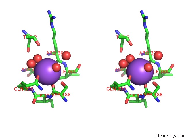
Stereo pair view
Mono view
Stereo pair view
A full contact list of Sodium with other atoms in the Na binding
site number 1 of Crystal Structure of A Glycoside Hydrolase Family 43 Arabinoxylan Arabinofuranohydrolase From Bacillus Subtilis in Complex with Cellotetraose. within 5.0Å range:
|
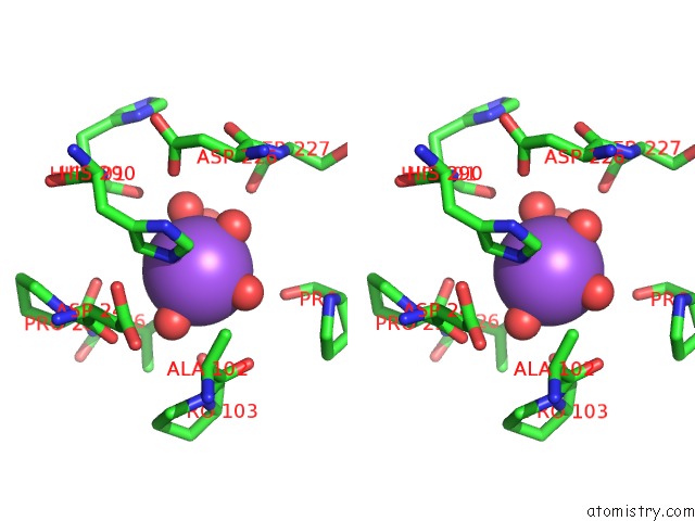
Sodium binding site 2 out of 2 in 3c7o
Go back to
Sodium binding site 2 out
of 2 in the Crystal Structure of A Glycoside Hydrolase Family 43 Arabinoxylan Arabinofuranohydrolase From Bacillus Subtilis in Complex with Cellotetraose.
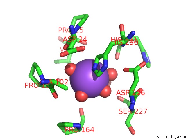
Mono view
Stereo pair view
Mono view
Stereo pair view
A full contact list of Sodium with other atoms in the Na binding
site number 2 of Crystal Structure of A Glycoside Hydrolase Family 43 Arabinoxylan Arabinofuranohydrolase From Bacillus Subtilis in Complex with Cellotetraose. within 5.0Å range:
|
Reference:
E.Vandermarliere,
T.M.Bourgois,
M.D.Winn,
S.Van Campenhout,
G.Volckaert,
J.A.Delcour,
S.V.Strelkov,
A.Rabijns,
C.M.Courtin.
Structural Analysis of A Glycoside Hydrolase Family 43 Arabinoxylan Arabinofuranohydrolase in Complex with Xylotetraose Reveals A Different Binding Mechanism Compared with Other Members of the Same Family. Biochem.J. V. 418 39 2009.
ISSN: ISSN 0264-6021
PubMed: 18980579
DOI: 10.1042/BJ20081256
Page generated: Mon Oct 7 06:13:22 2024
ISSN: ISSN 0264-6021
PubMed: 18980579
DOI: 10.1042/BJ20081256
Last articles
Cl in 5FJJCl in 5FJG
Cl in 5FHO
Cl in 5FJH
Cl in 5FJ3
Cl in 5FIP
Cl in 5FJ2
Cl in 5FIW
Cl in 5FIC
Cl in 5FHS