Sodium »
PDB 3bjp-3c7h »
3bk8 »
Sodium in PDB 3bk8: Urate Oxidase Aza-Xanthine Complex in Cyanide
Enzymatic activity of Urate Oxidase Aza-Xanthine Complex in Cyanide
All present enzymatic activity of Urate Oxidase Aza-Xanthine Complex in Cyanide:
1.7.3.3;
1.7.3.3;
Protein crystallography data
The structure of Urate Oxidase Aza-Xanthine Complex in Cyanide, PDB code: 3bk8
was solved by
L.Gabison,
T.Prange,
N.Colloc'h,
M.El Hajji,
B.Castro,
M.Chiadmi,
with X-Ray Crystallography technique. A brief refinement statistics is given in the table below:
| Resolution Low / High (Å) | 10.00 / 1.60 |
| Space group | I 2 2 2 |
| Cell size a, b, c (Å), α, β, γ (°) | 79.718, 95.105, 103.957, 90.00, 90.00, 90.00 |
| R / Rfree (%) | 20 / 23 |
Sodium Binding Sites:
The binding sites of Sodium atom in the Urate Oxidase Aza-Xanthine Complex in Cyanide
(pdb code 3bk8). This binding sites where shown within
5.0 Angstroms radius around Sodium atom.
In total only one binding site of Sodium was determined in the Urate Oxidase Aza-Xanthine Complex in Cyanide, PDB code: 3bk8:
In total only one binding site of Sodium was determined in the Urate Oxidase Aza-Xanthine Complex in Cyanide, PDB code: 3bk8:
Sodium binding site 1 out of 1 in 3bk8
Go back to
Sodium binding site 1 out
of 1 in the Urate Oxidase Aza-Xanthine Complex in Cyanide
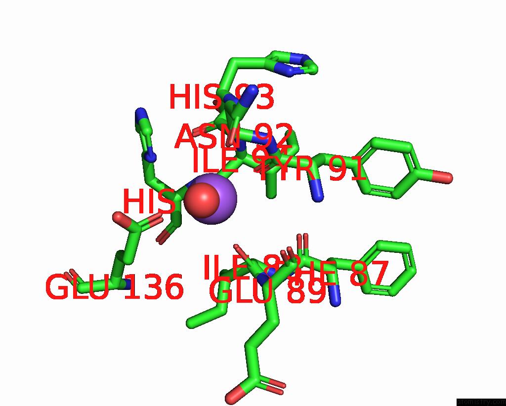
Mono view
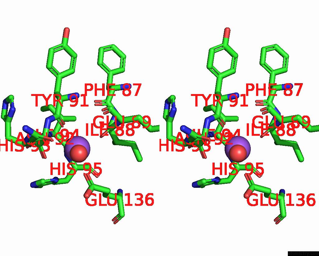
Stereo pair view
Mono view
Stereo pair view
A full contact list of Sodium with other atoms in the Na binding
site number 1 of Urate Oxidase Aza-Xanthine Complex in Cyanide within 5.0Å range:
|
Reference:
L.Gabison,
T.Prange,
N.Colloc'h,
M.El Hajji,
B.Castro,
M.Chiadmi.
Structural Analysis of Urate Oxidase in Complex with Its Natural Substrate Inhibited By Cyanide: Mechanistic Implications Bmc Struct.Biol. V. 8 32 2008.
ISSN: ESSN 1472-6807
PubMed: 18638417
DOI: 10.1186/1472-6807-8-32
Page generated: Mon Oct 7 06:05:17 2024
ISSN: ESSN 1472-6807
PubMed: 18638417
DOI: 10.1186/1472-6807-8-32
Last articles
Cl in 7XJBCl in 7XO8
Cl in 7XOV
Cl in 7XO7
Cl in 7XO9
Cl in 7XJO
Cl in 7XNC
Cl in 7XLP
Cl in 7XLI
Cl in 7XL9