Sodium »
PDB 3b2n-3biu »
3b72 »
Sodium in PDB 3b72: Crystal Structure of Lysozyme Folded in Sds and 2-Methyl-2,4- Pentanediol
Enzymatic activity of Crystal Structure of Lysozyme Folded in Sds and 2-Methyl-2,4- Pentanediol
All present enzymatic activity of Crystal Structure of Lysozyme Folded in Sds and 2-Methyl-2,4- Pentanediol:
3.2.1.17;
3.2.1.17;
Protein crystallography data
The structure of Crystal Structure of Lysozyme Folded in Sds and 2-Methyl-2,4- Pentanediol, PDB code: 3b72
was solved by
C.Michaux,
J.Pouyez,
J.Wouters,
G.G.Prive,
with X-Ray Crystallography technique. A brief refinement statistics is given in the table below:
| Resolution Low / High (Å) | 10.00 / 1.50 |
| Space group | P 43 21 2 |
| Cell size a, b, c (Å), α, β, γ (°) | 77.900, 77.900, 37.530, 90.00, 90.00, 90.00 |
| R / Rfree (%) | 20.4 / 24.1 |
Other elements in 3b72:
The structure of Crystal Structure of Lysozyme Folded in Sds and 2-Methyl-2,4- Pentanediol also contains other interesting chemical elements:
| Chlorine | (Cl) | 2 atoms |
Sodium Binding Sites:
The binding sites of Sodium atom in the Crystal Structure of Lysozyme Folded in Sds and 2-Methyl-2,4- Pentanediol
(pdb code 3b72). This binding sites where shown within
5.0 Angstroms radius around Sodium atom.
In total only one binding site of Sodium was determined in the Crystal Structure of Lysozyme Folded in Sds and 2-Methyl-2,4- Pentanediol, PDB code: 3b72:
In total only one binding site of Sodium was determined in the Crystal Structure of Lysozyme Folded in Sds and 2-Methyl-2,4- Pentanediol, PDB code: 3b72:
Sodium binding site 1 out of 1 in 3b72
Go back to
Sodium binding site 1 out
of 1 in the Crystal Structure of Lysozyme Folded in Sds and 2-Methyl-2,4- Pentanediol
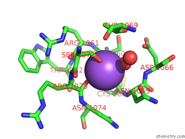
Mono view
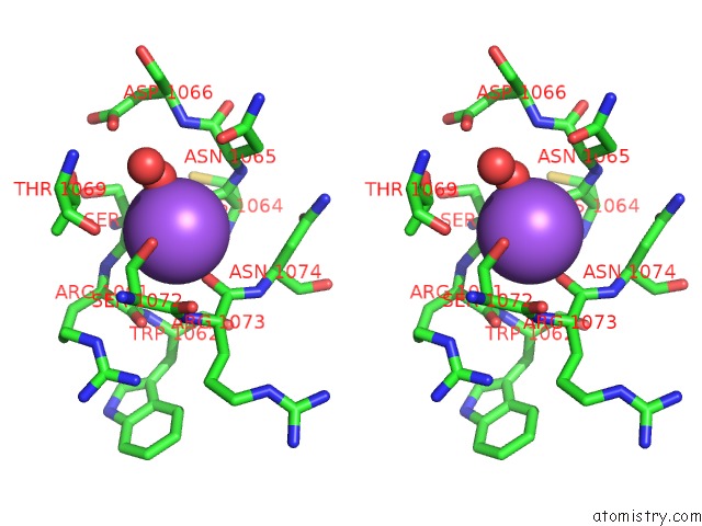
Stereo pair view
Mono view
Stereo pair view
A full contact list of Sodium with other atoms in the Na binding
site number 1 of Crystal Structure of Lysozyme Folded in Sds and 2-Methyl-2,4- Pentanediol within 5.0Å range:
|
Reference:
C.Michaux,
J.Pouyez,
J.Wouters,
G.G.Prive.
Protecting Role of Cosolvents in Protein Denaturation By Sds: A Structural Study. Bmc Struct.Biol. V. 8 29 2008.
ISSN: ESSN 1472-6807
PubMed: 18522744
DOI: 10.1186/1472-6807-8-29
Page generated: Mon Oct 7 06:00:04 2024
ISSN: ESSN 1472-6807
PubMed: 18522744
DOI: 10.1186/1472-6807-8-29
Last articles
Zn in 9J0NZn in 9J0O
Zn in 9J0P
Zn in 9FJX
Zn in 9EKB
Zn in 9C0F
Zn in 9CAH
Zn in 9CH0
Zn in 9CH3
Zn in 9CH1