Sodium »
PDB 3b2n-3biu »
3b3c »
Sodium in PDB 3b3c: Crystal Structure of the M180A Mutant of the Aminopeptidase From Vibrio Proteolyticus in Complex with Leucine Phosphonic Acid
Enzymatic activity of Crystal Structure of the M180A Mutant of the Aminopeptidase From Vibrio Proteolyticus in Complex with Leucine Phosphonic Acid
All present enzymatic activity of Crystal Structure of the M180A Mutant of the Aminopeptidase From Vibrio Proteolyticus in Complex with Leucine Phosphonic Acid:
3.4.11.10;
3.4.11.10;
Protein crystallography data
The structure of Crystal Structure of the M180A Mutant of the Aminopeptidase From Vibrio Proteolyticus in Complex with Leucine Phosphonic Acid, PDB code: 3b3c
was solved by
N.J.Ataie,
Q.Q.Hoang,
G.A.Petsko,
D.Ringe,
with X-Ray Crystallography technique. A brief refinement statistics is given in the table below:
| Resolution Low / High (Å) | 30.63 / 1.46 |
| Space group | P 61 2 2 |
| Cell size a, b, c (Å), α, β, γ (°) | 108.189, 108.189, 97.206, 90.00, 90.00, 120.00 |
| R / Rfree (%) | 19.7 / 22.6 |
Other elements in 3b3c:
The structure of Crystal Structure of the M180A Mutant of the Aminopeptidase From Vibrio Proteolyticus in Complex with Leucine Phosphonic Acid also contains other interesting chemical elements:
| Potassium | (K) | 1 atom |
| Zinc | (Zn) | 2 atoms |
Sodium Binding Sites:
The binding sites of Sodium atom in the Crystal Structure of the M180A Mutant of the Aminopeptidase From Vibrio Proteolyticus in Complex with Leucine Phosphonic Acid
(pdb code 3b3c). This binding sites where shown within
5.0 Angstroms radius around Sodium atom.
In total 6 binding sites of Sodium where determined in the Crystal Structure of the M180A Mutant of the Aminopeptidase From Vibrio Proteolyticus in Complex with Leucine Phosphonic Acid, PDB code: 3b3c:
Jump to Sodium binding site number: 1; 2; 3; 4; 5; 6;
In total 6 binding sites of Sodium where determined in the Crystal Structure of the M180A Mutant of the Aminopeptidase From Vibrio Proteolyticus in Complex with Leucine Phosphonic Acid, PDB code: 3b3c:
Jump to Sodium binding site number: 1; 2; 3; 4; 5; 6;
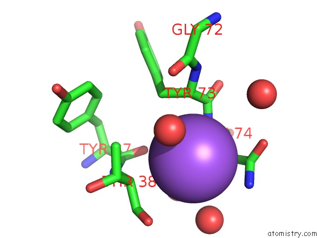
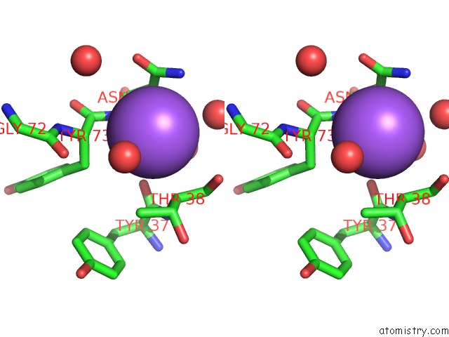
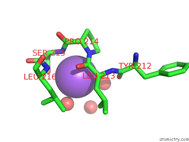
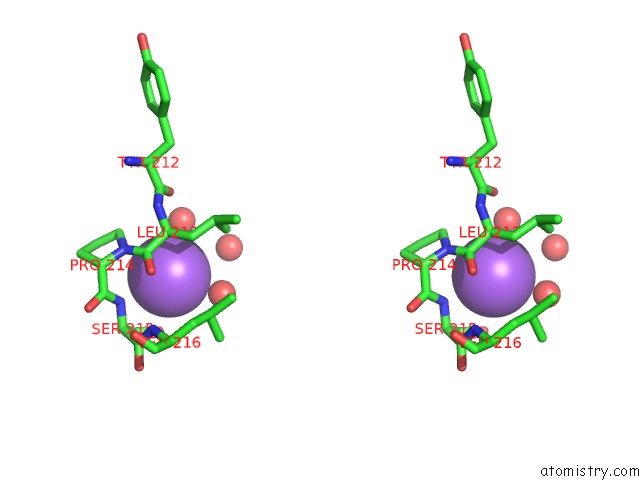
Sodium binding site 1 out of 6 in 3b3c
Go back to
Sodium binding site 1 out
of 6 in the Crystal Structure of the M180A Mutant of the Aminopeptidase From Vibrio Proteolyticus in Complex with Leucine Phosphonic Acid
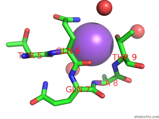
Mono view
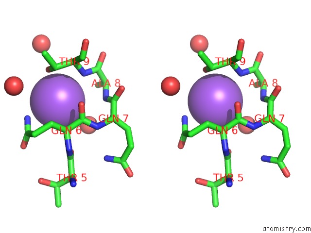
Stereo pair view
Mono view
Stereo pair view
A full contact list of Sodium with other atoms in the Na binding
site number 1 of Crystal Structure of the M180A Mutant of the Aminopeptidase From Vibrio Proteolyticus in Complex with Leucine Phosphonic Acid within 5.0Å range:
|
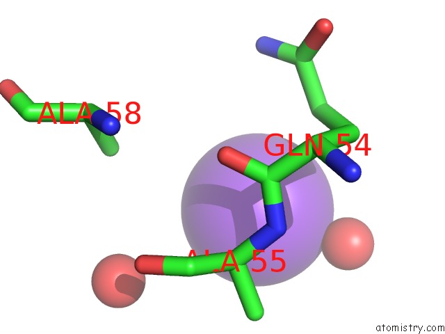
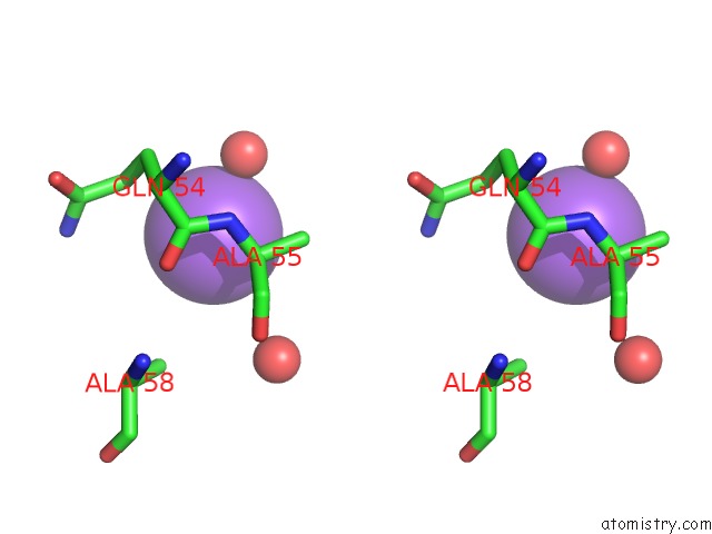
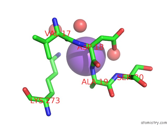
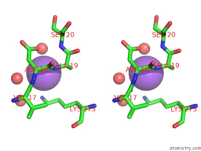
Sodium binding site 2 out of 6 in 3b3c
Go back to
Sodium binding site 2 out
of 6 in the Crystal Structure of the M180A Mutant of the Aminopeptidase From Vibrio Proteolyticus in Complex with Leucine Phosphonic Acid
Mono view
Stereo pair view
Mono view
Stereo pair view
A full contact list of Sodium with other atoms in the Na binding
site number 2 of Crystal Structure of the M180A Mutant of the Aminopeptidase From Vibrio Proteolyticus in Complex with Leucine Phosphonic Acid within 5.0Å range:
|
Sodium binding site 3 out of 6 in 3b3c
Go back to
Sodium binding site 3 out
of 6 in the Crystal Structure of the M180A Mutant of the Aminopeptidase From Vibrio Proteolyticus in Complex with Leucine Phosphonic Acid
Mono view
Stereo pair view
Mono view
Stereo pair view
A full contact list of Sodium with other atoms in the Na binding
site number 3 of Crystal Structure of the M180A Mutant of the Aminopeptidase From Vibrio Proteolyticus in Complex with Leucine Phosphonic Acid within 5.0Å range:
|
Sodium binding site 4 out of 6 in 3b3c
Go back to
Sodium binding site 4 out
of 6 in the Crystal Structure of the M180A Mutant of the Aminopeptidase From Vibrio Proteolyticus in Complex with Leucine Phosphonic Acid
Mono view
Stereo pair view
Mono view
Stereo pair view
A full contact list of Sodium with other atoms in the Na binding
site number 4 of Crystal Structure of the M180A Mutant of the Aminopeptidase From Vibrio Proteolyticus in Complex with Leucine Phosphonic Acid within 5.0Å range:
|
Sodium binding site 5 out of 6 in 3b3c
Go back to
Sodium binding site 5 out
of 6 in the Crystal Structure of the M180A Mutant of the Aminopeptidase From Vibrio Proteolyticus in Complex with Leucine Phosphonic Acid
Mono view
Stereo pair view
Mono view
Stereo pair view
A full contact list of Sodium with other atoms in the Na binding
site number 5 of Crystal Structure of the M180A Mutant of the Aminopeptidase From Vibrio Proteolyticus in Complex with Leucine Phosphonic Acid within 5.0Å range:
|
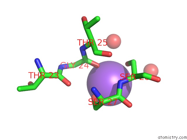
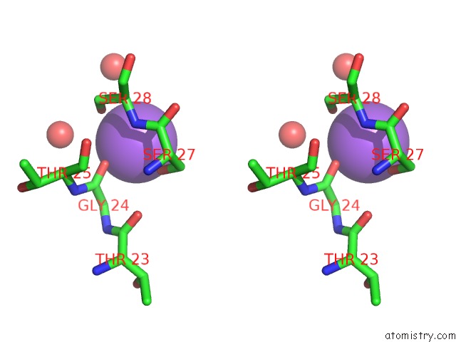
Sodium binding site 6 out of 6 in 3b3c
Go back to
Sodium binding site 6 out
of 6 in the Crystal Structure of the M180A Mutant of the Aminopeptidase From Vibrio Proteolyticus in Complex with Leucine Phosphonic Acid
Mono view
Stereo pair view
Mono view
Stereo pair view
A full contact list of Sodium with other atoms in the Na binding
site number 6 of Crystal Structure of the M180A Mutant of the Aminopeptidase From Vibrio Proteolyticus in Complex with Leucine Phosphonic Acid within 5.0Å range:
|
Reference:
N.J.Ataie,
Q.Q.Hoang,
M.P.Zahniser,
Y.Tu,
A.Milne,
G.A.Petsko,
D.Ringe.
Zinc Coordination Geometry and Ligand Binding Affinity: the Structural and Kinetic Analysis of the Second-Shell Serine 228 Residue and the Methionine 180 Residue of the Aminopeptidase From Vibrio Proteolyticus. Biochemistry V. 47 7673 2008.
ISSN: ISSN 0006-2960
PubMed: 18576673
DOI: 10.1021/BI702188E
Page generated: Mon Oct 7 05:58:24 2024
ISSN: ISSN 0006-2960
PubMed: 18576673
DOI: 10.1021/BI702188E
Last articles
Zn in 9J0NZn in 9J0O
Zn in 9J0P
Zn in 9FJX
Zn in 9EKB
Zn in 9C0F
Zn in 9CAH
Zn in 9CH0
Zn in 9CH3
Zn in 9CH1