Sodium »
PDB 3ajn-3b1q »
3b1e »
Sodium in PDB 3b1e: Crystal Structure of Betac-S Lyase From Streptococcus Anginosus in Complex with L-Serine: Alpha-Aminoacrylate Form
Enzymatic activity of Crystal Structure of Betac-S Lyase From Streptococcus Anginosus in Complex with L-Serine: Alpha-Aminoacrylate Form
All present enzymatic activity of Crystal Structure of Betac-S Lyase From Streptococcus Anginosus in Complex with L-Serine: Alpha-Aminoacrylate Form:
4.4.1.8;
4.4.1.8;
Protein crystallography data
The structure of Crystal Structure of Betac-S Lyase From Streptococcus Anginosus in Complex with L-Serine: Alpha-Aminoacrylate Form, PDB code: 3b1e
was solved by
Y.Kezuka,
Y.Yoshida,
T.Nonaka,
with X-Ray Crystallography technique. A brief refinement statistics is given in the table below:
| Resolution Low / High (Å) | 49.27 / 1.78 |
| Space group | P 21 21 21 |
| Cell size a, b, c (Å), α, β, γ (°) | 67.204, 111.212, 217.353, 90.00, 90.00, 90.00 |
| R / Rfree (%) | 14.2 / 17.7 |
Sodium Binding Sites:
The binding sites of Sodium atom in the Crystal Structure of Betac-S Lyase From Streptococcus Anginosus in Complex with L-Serine: Alpha-Aminoacrylate Form
(pdb code 3b1e). This binding sites where shown within
5.0 Angstroms radius around Sodium atom.
In total 4 binding sites of Sodium where determined in the Crystal Structure of Betac-S Lyase From Streptococcus Anginosus in Complex with L-Serine: Alpha-Aminoacrylate Form, PDB code: 3b1e:
Jump to Sodium binding site number: 1; 2; 3; 4;
In total 4 binding sites of Sodium where determined in the Crystal Structure of Betac-S Lyase From Streptococcus Anginosus in Complex with L-Serine: Alpha-Aminoacrylate Form, PDB code: 3b1e:
Jump to Sodium binding site number: 1; 2; 3; 4;
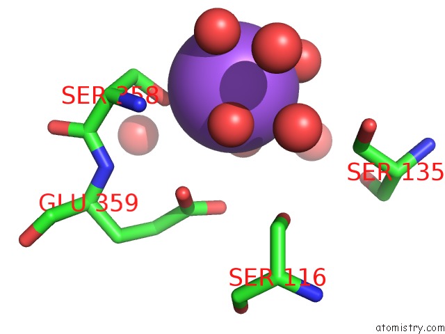
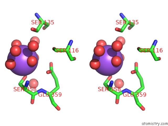
Sodium binding site 1 out of 4 in 3b1e
Go back to
Sodium binding site 1 out
of 4 in the Crystal Structure of Betac-S Lyase From Streptococcus Anginosus in Complex with L-Serine: Alpha-Aminoacrylate Form
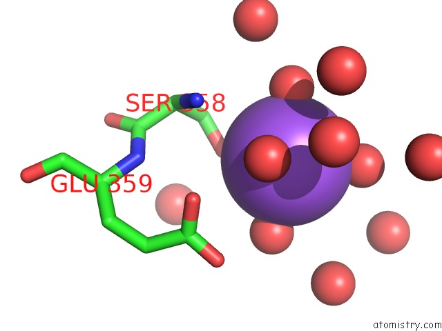
Mono view
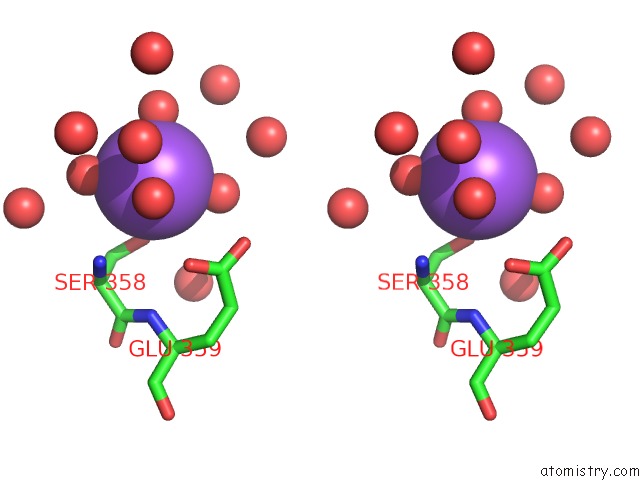
Stereo pair view
Mono view
Stereo pair view
A full contact list of Sodium with other atoms in the Na binding
site number 1 of Crystal Structure of Betac-S Lyase From Streptococcus Anginosus in Complex with L-Serine: Alpha-Aminoacrylate Form within 5.0Å range:
|
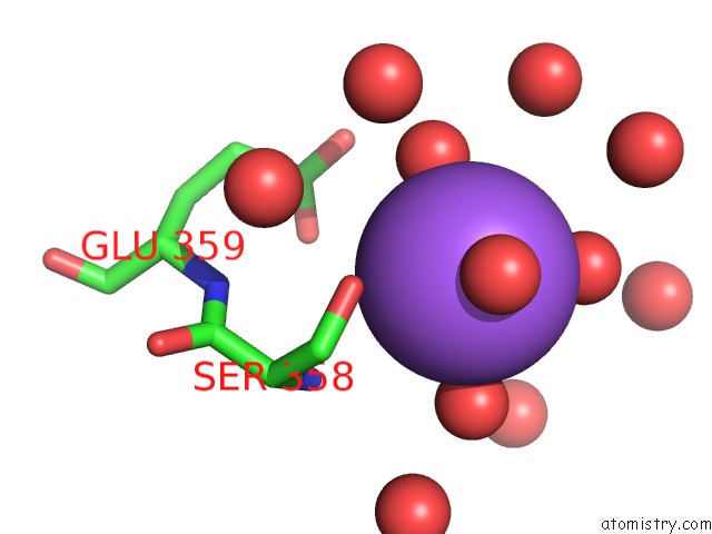
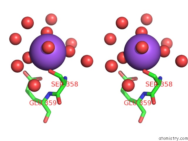
Sodium binding site 2 out of 4 in 3b1e
Go back to
Sodium binding site 2 out
of 4 in the Crystal Structure of Betac-S Lyase From Streptococcus Anginosus in Complex with L-Serine: Alpha-Aminoacrylate Form
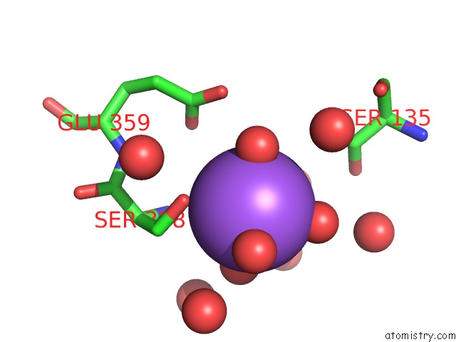
Mono view
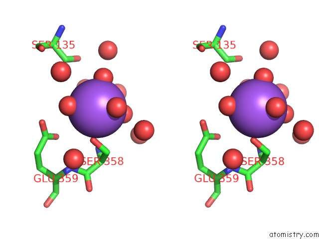
Stereo pair view
Mono view
Stereo pair view
A full contact list of Sodium with other atoms in the Na binding
site number 2 of Crystal Structure of Betac-S Lyase From Streptococcus Anginosus in Complex with L-Serine: Alpha-Aminoacrylate Form within 5.0Å range:
|
Sodium binding site 3 out of 4 in 3b1e
Go back to
Sodium binding site 3 out
of 4 in the Crystal Structure of Betac-S Lyase From Streptococcus Anginosus in Complex with L-Serine: Alpha-Aminoacrylate Form
Mono view
Stereo pair view
Mono view
Stereo pair view
A full contact list of Sodium with other atoms in the Na binding
site number 3 of Crystal Structure of Betac-S Lyase From Streptococcus Anginosus in Complex with L-Serine: Alpha-Aminoacrylate Form within 5.0Å range:
|
Sodium binding site 4 out of 4 in 3b1e
Go back to
Sodium binding site 4 out
of 4 in the Crystal Structure of Betac-S Lyase From Streptococcus Anginosus in Complex with L-Serine: Alpha-Aminoacrylate Form
Mono view
Stereo pair view
Mono view
Stereo pair view
A full contact list of Sodium with other atoms in the Na binding
site number 4 of Crystal Structure of Betac-S Lyase From Streptococcus Anginosus in Complex with L-Serine: Alpha-Aminoacrylate Form within 5.0Å range:
|
Reference:
Y.Kezuka,
Y.Yoshida,
T.Nonaka.
Structural Insights Into Catalysis By Beta C-S Lyase From Streptococcus Anginosus Proteins V. 80 2447 2012.
ISSN: ISSN 0887-3585
PubMed: 22674431
DOI: 10.1002/PROT.24129
Page generated: Mon Oct 7 05:54:52 2024
ISSN: ISSN 0887-3585
PubMed: 22674431
DOI: 10.1002/PROT.24129
Last articles
Cl in 7TLGCl in 7TJC
Cl in 7TJO
Cl in 7TLE
Cl in 7TKV
Cl in 7THH
Cl in 7TIV
Cl in 7TIW
Cl in 7TI9
Cl in 7TIU