Sodium »
PDB 3ajn-3b1q »
3ass »
Sodium in PDB 3ass: Crystal Structure of P Domain From Norovirus FUNABASHI258 Stain in the Complex with Lewis-B
Protein crystallography data
The structure of Crystal Structure of P Domain From Norovirus FUNABASHI258 Stain in the Complex with Lewis-B, PDB code: 3ass
was solved by
T.Kubota,
A.Kumagai,
H.Itoh,
S.Furukawa,
H.Narimatsu,
T.Wakita,
K.Ishii,
N.Takeda,
Y.Someya,
H.Shirato,
with X-Ray Crystallography technique. A brief refinement statistics is given in the table below:
| Resolution Low / High (Å) | 31.23 / 1.60 |
| Space group | P 31 |
| Cell size a, b, c (Å), α, β, γ (°) | 74.690, 74.690, 106.973, 90.00, 90.00, 120.00 |
| R / Rfree (%) | 18.5 / 20.2 |
Sodium Binding Sites:
The binding sites of Sodium atom in the Crystal Structure of P Domain From Norovirus FUNABASHI258 Stain in the Complex with Lewis-B
(pdb code 3ass). This binding sites where shown within
5.0 Angstroms radius around Sodium atom.
In total 2 binding sites of Sodium where determined in the Crystal Structure of P Domain From Norovirus FUNABASHI258 Stain in the Complex with Lewis-B, PDB code: 3ass:
Jump to Sodium binding site number: 1; 2;
In total 2 binding sites of Sodium where determined in the Crystal Structure of P Domain From Norovirus FUNABASHI258 Stain in the Complex with Lewis-B, PDB code: 3ass:
Jump to Sodium binding site number: 1; 2;
Sodium binding site 1 out of 2 in 3ass
Go back to
Sodium binding site 1 out
of 2 in the Crystal Structure of P Domain From Norovirus FUNABASHI258 Stain in the Complex with Lewis-B
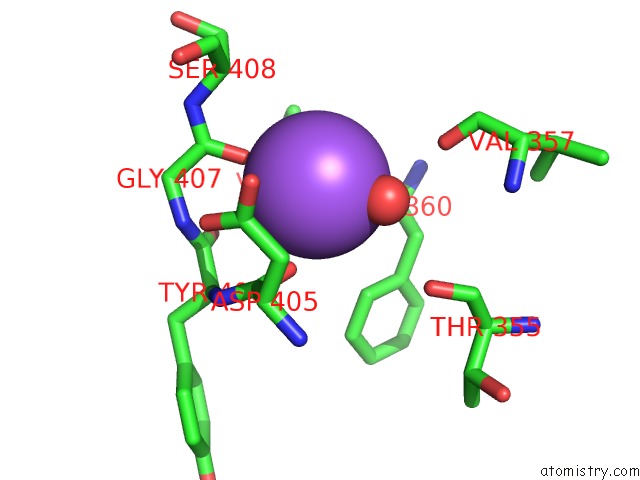
Mono view
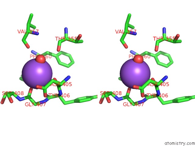
Stereo pair view
Mono view
Stereo pair view
A full contact list of Sodium with other atoms in the Na binding
site number 1 of Crystal Structure of P Domain From Norovirus FUNABASHI258 Stain in the Complex with Lewis-B within 5.0Å range:
|
Sodium binding site 2 out of 2 in 3ass
Go back to
Sodium binding site 2 out
of 2 in the Crystal Structure of P Domain From Norovirus FUNABASHI258 Stain in the Complex with Lewis-B
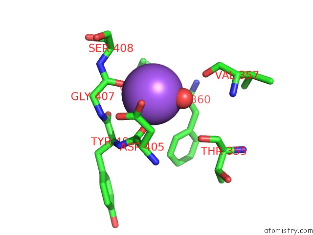
Mono view
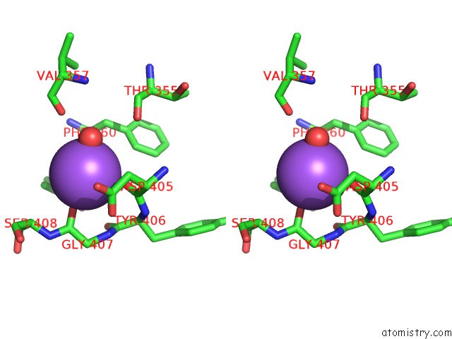
Stereo pair view
Mono view
Stereo pair view
A full contact list of Sodium with other atoms in the Na binding
site number 2 of Crystal Structure of P Domain From Norovirus FUNABASHI258 Stain in the Complex with Lewis-B within 5.0Å range:
|
Reference:
T.Kubota,
A.Kumagai,
H.Ito,
S.Furukawa,
Y.Someya,
N.Takeda,
K.Ishii,
T.Wakita,
H.Narimatsu,
H.Shirato.
Structural Basis For the Recognition of Lewis Antigens By Genogroup I Norovirus J.Virol. V. 86 11138 2012.
ISSN: ISSN 0022-538X
PubMed: 22855491
DOI: 10.1128/JVI.00278-12
Page generated: Mon Oct 7 05:52:20 2024
ISSN: ISSN 0022-538X
PubMed: 22855491
DOI: 10.1128/JVI.00278-12
Last articles
Zn in 9J0NZn in 9J0O
Zn in 9J0P
Zn in 9FJX
Zn in 9EKB
Zn in 9C0F
Zn in 9CAH
Zn in 9CH0
Zn in 9CH3
Zn in 9CH1