Sodium »
PDB 3ajn-3b1q »
3aki »
Sodium in PDB 3aki: Crystal Structure of Exo-1,5-Alpha-L-Arabinofuranosidase Complexed with Alpha-L-Arabinofuranosyl Azido
Enzymatic activity of Crystal Structure of Exo-1,5-Alpha-L-Arabinofuranosidase Complexed with Alpha-L-Arabinofuranosyl Azido
All present enzymatic activity of Crystal Structure of Exo-1,5-Alpha-L-Arabinofuranosidase Complexed with Alpha-L-Arabinofuranosyl Azido:
3.2.1.55;
3.2.1.55;
Protein crystallography data
The structure of Crystal Structure of Exo-1,5-Alpha-L-Arabinofuranosidase Complexed with Alpha-L-Arabinofuranosyl Azido, PDB code: 3aki
was solved by
Z.Fujimoto,
H.Ichinose,
S.Kaneko,
with X-Ray Crystallography technique. A brief refinement statistics is given in the table below:
| Resolution Low / High (Å) | 74.79 / 2.00 |
| Space group | P 21 21 21 |
| Cell size a, b, c (Å), α, β, γ (°) | 41.026, 89.719, 135.424, 90.00, 90.00, 90.00 |
| R / Rfree (%) | 19.3 / 23.1 |
Other elements in 3aki:
The structure of Crystal Structure of Exo-1,5-Alpha-L-Arabinofuranosidase Complexed with Alpha-L-Arabinofuranosyl Azido also contains other interesting chemical elements:
| Chlorine | (Cl) | 1 atom |
Sodium Binding Sites:
The binding sites of Sodium atom in the Crystal Structure of Exo-1,5-Alpha-L-Arabinofuranosidase Complexed with Alpha-L-Arabinofuranosyl Azido
(pdb code 3aki). This binding sites where shown within
5.0 Angstroms radius around Sodium atom.
In total only one binding site of Sodium was determined in the Crystal Structure of Exo-1,5-Alpha-L-Arabinofuranosidase Complexed with Alpha-L-Arabinofuranosyl Azido, PDB code: 3aki:
In total only one binding site of Sodium was determined in the Crystal Structure of Exo-1,5-Alpha-L-Arabinofuranosidase Complexed with Alpha-L-Arabinofuranosyl Azido, PDB code: 3aki:
Sodium binding site 1 out of 1 in 3aki
Go back to
Sodium binding site 1 out
of 1 in the Crystal Structure of Exo-1,5-Alpha-L-Arabinofuranosidase Complexed with Alpha-L-Arabinofuranosyl Azido
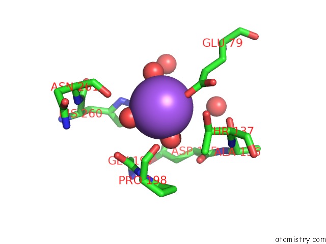
Mono view
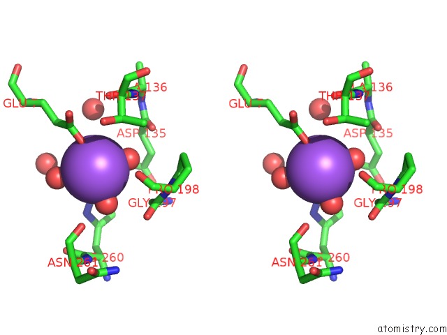
Stereo pair view
Mono view
Stereo pair view
A full contact list of Sodium with other atoms in the Na binding
site number 1 of Crystal Structure of Exo-1,5-Alpha-L-Arabinofuranosidase Complexed with Alpha-L-Arabinofuranosyl Azido within 5.0Å range:
|
Reference:
Z.Fujimoto,
H.Ichinose,
T.Maehara,
M.Honda,
M.Kitaoka,
S.Kaneko.
Crystal Structure of An Exo-1,5-{Alpha}-L-Arabinofuranosidase From Streptomyces Avermitilis Provides Insights Into the Mechanism of Substrate Discrimination Between Exo- and Endo-Type Enzymes in Glycoside Hydrolase Family 43. J.Biol.Chem. V. 285 34134 2010.
ISSN: ISSN 0021-9258
PubMed: 20739278
DOI: 10.1074/JBC.M110.164251
Page generated: Mon Oct 7 05:50:28 2024
ISSN: ISSN 0021-9258
PubMed: 20739278
DOI: 10.1074/JBC.M110.164251
Last articles
Cl in 5I6XCl in 5I5Y
Cl in 5I6U
Cl in 5I60
Cl in 5I5W
Cl in 5I5T
Cl in 5I5S
Cl in 5I5V
Cl in 5I5U
Cl in 5I58