Sodium »
PDB 2yfa-2zgb »
2z5d »
Sodium in PDB 2z5d: Human Ubiquitin-Conjugating Enzyme E2 H
Enzymatic activity of Human Ubiquitin-Conjugating Enzyme E2 H
All present enzymatic activity of Human Ubiquitin-Conjugating Enzyme E2 H:
6.3.2.19;
6.3.2.19;
Protein crystallography data
The structure of Human Ubiquitin-Conjugating Enzyme E2 H, PDB code: 2z5d
was solved by
A.Bochkarev,
H.Cui,
J.R.Walker,
E.M.Newman,
F.Mackenzie,
K.P.Battaile,
M.Sundstrom,
C.Arrowsmith,
A.Edwards,
S.Dhe-Paganon,
Structural Genomicsconsortium (Sgc),
with X-Ray Crystallography technique. A brief refinement statistics is given in the table below:
| Resolution Low / High (Å) | 65.80 / 2.10 |
| Space group | P 21 21 21 |
| Cell size a, b, c (Å), α, β, γ (°) | 42.959, 85.861, 102.558, 90.00, 90.00, 90.00 |
| R / Rfree (%) | 18.1 / 22.3 |
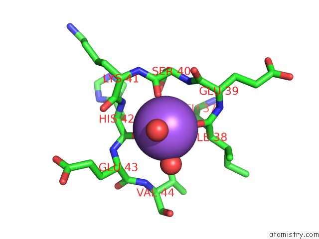
Sodium Binding Sites:
The binding sites of Sodium atom in the Human Ubiquitin-Conjugating Enzyme E2 H
(pdb code 2z5d). This binding sites where shown within
5.0 Angstroms radius around Sodium atom.
In total only one binding site of Sodium was determined in the Human Ubiquitin-Conjugating Enzyme E2 H, PDB code: 2z5d:
In total only one binding site of Sodium was determined in the Human Ubiquitin-Conjugating Enzyme E2 H, PDB code: 2z5d:
Sodium binding site 1 out of 1 in 2z5d
Go back to
Sodium binding site 1 out
of 1 in the Human Ubiquitin-Conjugating Enzyme E2 H
Mono view
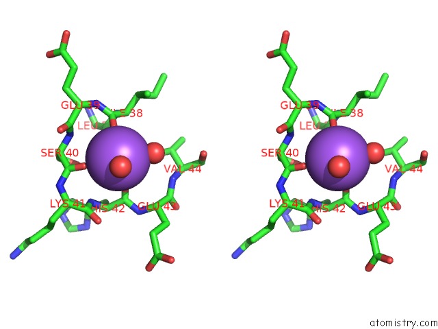
Stereo pair view
Mono view
Stereo pair view
A full contact list of Sodium with other atoms in the Na binding
site number 1 of Human Ubiquitin-Conjugating Enzyme E2 H within 5.0Å range:
|
Reference:
Y.Sheng,
J.H.Hong,
R.Doherty,
T.Srikumar,
J.Shloush,
G.V.Avvakumov,
J.R.Walker,
S.Xue,
D.Neculai,
J.W.Wan,
S.K.Kim,
C.H.Arrowsmith,
B.Raught,
S.Dhe-Paganon.
A Human Ubiquitin Conjugating Enzyme (E2)-Hect E3 Ligase Structure-Function Screen. Mol.Cell Proteomics V. 11 329 2012.
ISSN: ISSN 1535-9476
PubMed: 22496338
DOI: 10.1074/MCP.O111.013706
Page generated: Mon Oct 7 05:30:19 2024
ISSN: ISSN 1535-9476
PubMed: 22496338
DOI: 10.1074/MCP.O111.013706
Last articles
Cl in 5J2BCl in 5J2A
Cl in 5IXL
Cl in 5J0Z
Cl in 5J1T
Cl in 5J1S
Cl in 5IXY
Cl in 5IXE
Cl in 5IXS
Cl in 5IVT