Sodium »
PDB 2yfa-2zgb »
2yyk »
Sodium in PDB 2yyk: Crystal Structure of the Mutant of Hpab (T198I, A276G, and R466H)
Enzymatic activity of Crystal Structure of the Mutant of Hpab (T198I, A276G, and R466H)
All present enzymatic activity of Crystal Structure of the Mutant of Hpab (T198I, A276G, and R466H):
1.14.13.3;
1.14.13.3;
Protein crystallography data
The structure of Crystal Structure of the Mutant of Hpab (T198I, A276G, and R466H), PDB code: 2yyk
was solved by
S.-H.Kim,
T.Hisano,
K.Takeda,
W.Iwasaki,
A.Ebihara,
K.Miki,
with X-Ray Crystallography technique. A brief refinement statistics is given in the table below:
| Resolution Low / High (Å) | 41.53 / 1.60 |
| Space group | I 2 2 2 |
| Cell size a, b, c (Å), α, β, γ (°) | 91.819, 99.612, 131.091, 90.00, 90.00, 90.00 |
| R / Rfree (%) | 19.5 / 20.3 |
Sodium Binding Sites:
The binding sites of Sodium atom in the Crystal Structure of the Mutant of Hpab (T198I, A276G, and R466H)
(pdb code 2yyk). This binding sites where shown within
5.0 Angstroms radius around Sodium atom.
In total only one binding site of Sodium was determined in the Crystal Structure of the Mutant of Hpab (T198I, A276G, and R466H), PDB code: 2yyk:
In total only one binding site of Sodium was determined in the Crystal Structure of the Mutant of Hpab (T198I, A276G, and R466H), PDB code: 2yyk:
Sodium binding site 1 out of 1 in 2yyk
Go back to
Sodium binding site 1 out
of 1 in the Crystal Structure of the Mutant of Hpab (T198I, A276G, and R466H)
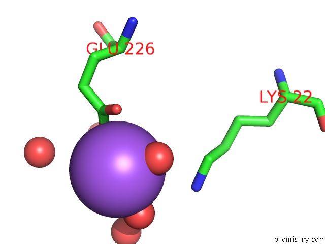
Mono view
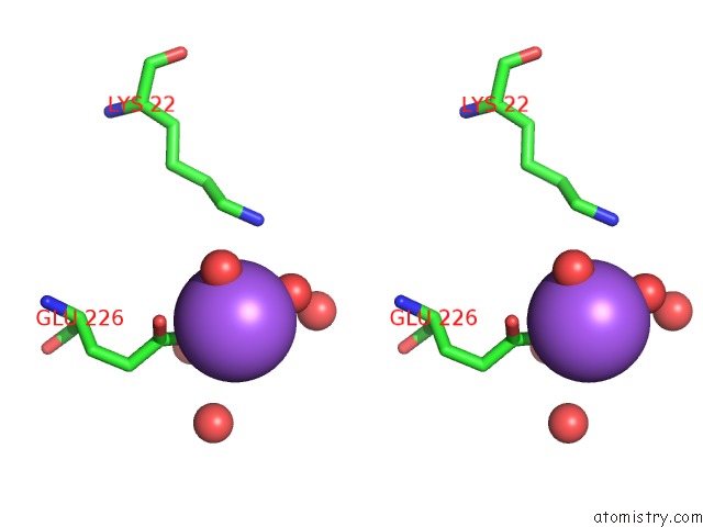
Stereo pair view
Mono view
Stereo pair view
A full contact list of Sodium with other atoms in the Na binding
site number 1 of Crystal Structure of the Mutant of Hpab (T198I, A276G, and R466H) within 5.0Å range:
|
Reference:
S.-H.Kim,
T.Hisano,
K.Takeda,
W.Iwasaki,
A.Ebihara,
K.Miki.
Crystal Structure of the Oxygenase Component (Hpab) of the 4-Hydroxyphenylacetate 3-Monooxygenase From Thermus Thermophilus HB8 J.Biol.Chem. V. 282 33107 2007.
ISSN: ISSN 0021-9258
PubMed: 17804419
DOI: 10.1074/JBC.M703440200
Page generated: Mon Oct 7 05:28:30 2024
ISSN: ISSN 0021-9258
PubMed: 17804419
DOI: 10.1074/JBC.M703440200
Last articles
Zn in 9MJ5Zn in 9HNW
Zn in 9G0L
Zn in 9FNE
Zn in 9DZN
Zn in 9E0I
Zn in 9D32
Zn in 9DAK
Zn in 8ZXC
Zn in 8ZUF